
MARPE Consultation Workflow:
Records, CBCT,
and Diagnostic Staging in One Appointment
A step-by-step framework for orthodontists to sequence the clinical exam, cone-beam CT, and suture-maturity assessment before leaving the chair.
TL;DR The MARPE consultation workflow integrates clinical records, CBCT acquisition, and suture-maturity staging into a single appointment to prevent diagnostic gaps that delay treatment. Sequencing the physical exam before CBCT acquisition improves scan prescription accuracy. Evidence from Chun et al. (2022) confirms CBCT imaging at baseline and post-expansion as the diagnostic standard for tracking midpalatal suture response.
Structuring a single-visit diagnostic workup around a coherent MARPE consultation workflow is one of the more consequential workflow decisions an orthodontist can make. A disorganized sequence—records one day, CBCT another, diagnostic discussion a third—introduces interpretation lag, risks missed candidacy signals, and erodes patient confidence in the planning process. Dr. Mark Radzhabov at ortodontmark.com has developed a sequenced, evidence-driven protocol that moves from clinical examination through CBCT acquisition to suture-maturity staging without leaving the chair. This article outlines that framework for orthodontists seeking a clinically practical, defensible consultation structure.
Why Does Appointment Sequencing Change
Diagnostic Accuracy
in Palatal Expansion Planning?
The MARPE consultation workflow is a sequenced single-visit protocol that integrates transverse clinical examination, CBCT acquisition with defined imaging criteria, and midpalatal suture-maturity staging into one diagnostic appointment before any appliance discussion begins. When records are collected across separate visits, clinicians frequently prescribe CBCT scans without the benefit of a prior clinical transverse assessment—resulting in scan fields that miss critical anatomy or fail to capture bilateral suture symmetry at the anterior palate. Clinical examination should precede CBCT prescription in every MARPE consultation. Palpation of the midpalatal raphe, assessment of buccal corridors, and measurement of inter-molar and inter-premolar widths on dental casts establish the magnitude of transverse deficiency and guide whether the CBCT field of view must extend to the nasal floor and greater palatine foramina. Chun et al. (2022) demonstrated that MARPE produced greater skeletal expansion of nasal width and increased greater palatine foramen dimensions compared to RPE, confirming that these anatomical landmarks are diagnostically relevant and must be visible on the scan. Age and growth status inform CBCT slice selection but do not replace it. Kinzinger et al. (2022) showed that expansion patterns differ significantly by age—parallel in children under 10 years and V-shaped in adolescents 12 years and older—meaning the coronal slice orientation chosen for suture assessment must account for the likely expansion geometry before the clinician ever places a region-of-interest cursor. Locking in the right imaging parameters during the clinical exam phase, not retrospectively, is what makes single-visit staging achievable. For a deeper review of miniscrew-assisted expansion planning protocol, the full framework is available at ortodontmark.com.
How Should You Read the Midpalatal Suture
Before Selecting
Between MARPE and Conventional RPE?
Once CBCT is acquired, the diagnostic discussion moves to suture-maturity staging in the same appointment. The coronal and axial reconstructions should be reviewed at consistent window-and-level settings across the anterior, middle, and posterior thirds of the palate. A mature, interdigitated suture presents as irregular cortical bridging rather than the clean radiolucent line seen in younger patients—and this morphology is the primary criterion separating MARPE candidates from those who require surgical assistance. Chun et al. (2022) randomized 40 patients with a mean age of approximately 14 years into equal groups of 20 and confirmed that midpalatal suture separation occurred in 95% of MARPE patients, with MARPE producing significantly greater maxillary width at both premolar and molar regions compared to RPE. The implication for the consultation is direct: if the suture morphology on CBCT is consistent with the patient population in that trial, the clinician can proceed with MARPE candidacy confidence rather than deferring to a second-opinion scan. Expansion force magnitude is a parallel consideration during the same diagnostic discussion. Camporesi et al. (2013) found that Hyrax and A2620 screws generated over 20 kg of force while Palatal Split screws generated approximately 16 kg, and that RPE appliances typically produce expansion forces ranging from 7.54 to 15.8 kg. Mapping the patient's suture density to an appropriate force range—rather than defaulting to a single protocol—is a staging decision that must happen before appliance selection, not after.
When Does the Chairside Diagnostic Discussion
Change
the Treatment Plan in Real Time?
The third phase of the single-visit diagnostic workup is the diagnostic discussion itself—conducted with the CBCT reconstructions displayed, dental casts in hand, and the clinical exam findings fresh. This sequencing matters because it allows the clinician to anchor every recommendation to an observable finding rather than a general protocol statement. Pointing to suture interdigitation on the axial slice while explaining the force threshold decision is materially more persuasive than describing the procedure abstractly. Chun et al. (2022) also documented that MARPE resulted in significantly less buccal tipping of anchor teeth during expansion and consolidation compared to RPE. This finding translates directly into counseling language: when the CBCT shows adequate bone volume lateral to the midpalate for miniscrew placement, the clinician can explain that the bone-borne force path reduces the dental compensations that complicate later alignment. The discussion becomes specific to that patient's imaging, not generic. Kinzinger et al. (2022) demonstrated that expansion patterns in children under 10 years follow a parallel vector, while adolescents 12 years and older show V-shaped opening. Sharing this age-related geometry during the diagnostic discussion helps the family understand why the same appliance behaves differently across siblings, and why CBCT-guided staging rather than chronological age determines the protocol. Orthodontists who want to see how this diagnostic flow integrates into a full clinical framework can submit a case for consultation at ortodontmark.com.
Free Clinical Guide
Which Expansion Appliance at Which Age?
Get Dr. Mark Radzhabov's guide to skeletal-expansion appliances — indications, pros & cons, and how to choose by patient age. Free to your inbox.
Prefer video? Watch the free demo lesson on MARPE appliance types, features and trade-offs.
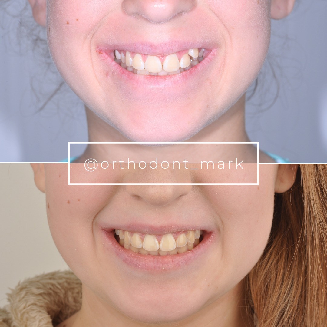
Documented outcomes
Clinical cases from Dr. Mark Radzhabov's practice


Learn the full MARPE protocol from Dr. Mark Radzhabov
Fundamental course covering CBCT patient selection, miniscrew planning, activation protocols, and 60+ clinical cases. Choose the access level that fits your practice.
Essentials of rapid palatal expansion for practicing orthodontists.
- Core RPE concepts and biomechanics
- 6 structured video lessons
- Clinical decision checklists
- Lifetime access to recordings
Deep-dive into MARPE protocol, diagnostics, and clinical execution.
- 6 modules covering MARPE from A to Z
- CBCT case selection walkthroughs
- Miniscrew placement and activation
- 60+ annotated clinical cases
- Direct Q&A with Dr. Mark Radzhabov
5-element medical consultation framework for dentists and orthodontists.
- Trust-building consultation protocol
- 5 lesson modules
- Templates for treatment plan delivery
- Works with any clinical specialty
Clinical FAQ
How should the MARPE consultation workflow be sequenced to avoid missing suture-maturity findings?
Complete the clinical transverse exam and dental-cast measurement first, then prescribe CBCT with those findings guiding field-of-view parameters. Review suture morphology and stage maturity before any appliance discussion. This three-step sequence prevents diagnostic gaps that force a second visit.
What CBCT imaging criteria should be specified before acquiring a palatal expansion scan?
Specify bilateral coverage from the nasal floor to the alveolar crest, include the greater palatine foramina, and use slice thickness appropriate for suture-margin resolution. Chun et al. (2022) used CBCT at baseline and post-expansion to capture both nasal width and suture separation changes.
At what point in the single-visit diagnostic workup should appliance selection be discussed?
Only after CBCT suture staging is complete. Suture morphology determines whether bone-borne MARPE or conventional RPE is appropriate. Chun et al. (2022) confirmed that MARPE produced significantly greater maxillary width and less anchor-tooth tipping—findings that should be visible on screen before the clinician recommends an appliance.
How does age affect expansion geometry in the CBCT diagnostic discussion?
Kinzinger et al. (2022) showed expansion patterns are parallel in children under 10 years and V-shaped in adolescents 12 years and older. Identifying which geometry applies to your patient informs coronal slice orientation and helps calibrate post-expansion CBCT comparisons.
Which screw force outputs should inform appliance selection during the MARPE diagnostic discussion?
Camporesi et al. (2013) found Hyrax and A2620 screws exceeded 20 kg of force while Palatal Split screws generated approximately 16 kg. RPE appliances broadly produce forces from 7.54 to 15.8 kg. Match screw selection to the resistance profile indicated by the patient's suture density on CBCT.
What anatomical landmarks must be visible on the CBCT to complete a chairside MARPE candidacy assessment?
The midpalatal suture in axial and coronal planes, the nasal floor, and bilateral greater palatine foramina. Chun et al. (2022) documented that MARPE produced greater nasal width and increased greater palatine foramen dimensions, confirming these landmarks carry diagnostic weight beyond suture staging alone.
How does buccal tipping risk factor into the diagnostic records workflow for MARPE versus RPE?
Chun et al. (2022) confirmed significantly less buccal tipping of anchor teeth with MARPE compared to RPE during both expansion and consolidation. When CBCT shows adequate palatal bone volume for miniscrew placement, documenting this advantage during the consultation supports the bone-borne treatment recommendation.
What suture separation rates should a clinician cite when counseling a patient during the diagnostic discussion?
Chun et al. (2022) reported midpalatal suture separation in 95% of MARPE patients and 90% of RPE patients in a group of 40 patients with a mean age of approximately 14 years. These figures apply when the patient population and suture maturity are comparable to that trial's inclusion criteria.
How many CBCT timepoints are standard in an evidence-based rapid palatal expansion diagnostic protocol?
Chun et al. (2022) used three timepoints—baseline, immediately after 35 turns of expansion, and after a 3-month consolidation period. The baseline scan acquired at the consultation appointment is the first of these three and sets the reference against which suture separation and nasal change are measured.
How does a structured MARPE training course help orthodontists implement single-visit diagnostic workflows?
Structured MARPE training walks through examination sequencing, CBCT prescription criteria, and suture-staging interpretation with case-based imaging examples. This closes the gap between published evidence and chairside execution—allowing orthodontists to complete candidacy assessment, imaging review, and diagnostic discussion within a single appointment.
A well-structured MARPE consultation workflow compresses what can otherwise be a multi-visit process into a single high-yield appointment—capturing the clinical exam, CBCT, and diagnostic staging before the patient leaves the room. The difference between a successful split and an avoidable failure frequently traces back to how thoroughly candidacy was assessed at the outset, not to the mechanics of the appliance itself. Orthodontists looking to implement or refine this protocol are encouraged to review Dr. Mark Radzhabov's structured MARPE training resources at ortodontmark.com or submit a case for direct consultation. Key sources: Chun et al. (2022), BMC Oral Health, doi:https://doi.org/10.1186/s12903–022-02138-w. Kinzinger et al. (2022), J Orofac Orthop, doi:https://doi.org/10.1007/s00056–022-00429-z. Camporesi et al. (2013), BioMedical Engineering OnLine, doi:http://www.biomedical-engineering-online.com/content/12/1/128.

