
When to Refer MARPE Case Review:
Referral
Thresholds and Multi-Provider Handoff
Evidence-based scope boundaries and imaging criteria that tell you when independent MARPE planning ends and specialist review begins.
TL;DR When to refer MARPE case review is a decision shaped by suture maturation staging, transverse deficiency severity, and provider scope—not patient age alone. Govaerts et al. (2023) found that by age 15, 61% of females already show fused midpalatal sutures at stages D and E, making timely CBCT triage and structured multi-provider handoff protocols essential for safe case selection.
A general orthodontist encounters a posterior crossbite, orders a CBCT, and faces a midpalatal suture that no longer resembles the textbook image from residency. Knowing precisely when to refer a MARPE case review—rather than proceeding independently—is one of the highest-stakes triage decisions in contemporary expansion practice. Dr. Mark Radzhabov at ortodontmark.com has structured this clinical guide around peer-reviewed imaging evidence to give orthodontists clear referral thresholds, scope boundaries, and handoff checkpoints before committing a patient to bone-borne expansion or surgical escalation.
What Does a Multi-Provider MARPE
Handoff
Actually Require at the Case Level?
A multi-provider MARPE handoff is a structured clinical transfer in which the referring orthodontist documents suture maturation stage, transverse deficiency magnitude, and provider-specific scope limits before a specialist assumes responsibility for expansion planning or surgical triage. Without this documentation layer, responsibility gaps appear between the imaging interpretation, the miniscrew placement, and the activation protocol—three tasks that do not always sit with the same clinician. The handoff is triggered by case complexity, not by appliance type alone. A tooth-borne rapid palatal expander in a young adolescent sits within most general orthodontic scopes. Once bone-borne anchorage is required—with titanium miniscrews engaging the palatal cortex between the first premolars and first molars—the procedural and radiographic demands escalate. Chun et al. (2022) demonstrated in a randomized clinical trial that MARPE produced significantly greater maxillary width gains at premolar and molar regions compared to conventional RPE (p < 0.05), confirming that the skeletal effect is meaningfully different and that the clinical stakes of incorrect device selection are proportionally higher. Referral thresholds should therefore be defined in advance within any multi-provider network. The referring clinician's scope covers diagnosis, CBCT acquisition, suture staging, and transverse discrepancy quantification. The specialist's scope covers miniscrew placement planning, surgical escalation decisions, and activation management. Documenting this boundary in the patient record—before treatment begins—is what transforms an informal referral into a defensible handoff. For a deeper review of appliance biomechanics that inform these scope lines, explore MARPE case selection and appliance protocols at ortodontmark.com.
Which CBCT Findings Should Immediately
Trigger
a Specialist Referral Before MARPE?
Midpalatal suture maturation staging on CBCT is the primary gating criterion for every expansion referral decision. A fused or near-fused suture identified in the anterior, middle, and posterior thirds—appearing as continuous cortical bridging rather than a radiolucent separation line—signals that conventional MARPE force delivery is unlikely to produce reliable sutural opening without surgical preparation. Govaerts et al. (2023) documented in a CBCT retrospective cohort study that at age 15 in females, 61% already show closed midpalatal sutures at stages D and E, a finding that directly challenges any age-based referral shortcut. Adjacent suture data sharpens the referral picture further. Govaerts et al. (2023) reported that pterygomaxillary suture closure reaches 83–100% and transpalatal suture closure reaches 78–85% by age 15 in females aged 13–17 years. A case presenting with concurrent pterygomaxillary and transpalatal closure alongside a stage D or E midpalatal suture represents a surgical triage scenario, not a MARPE-first scenario, and the referral to a maxillofacial surgeon should occur before any activation plan is written. Expert consensus reported by Govaerts et al. (2023) placed the surgical RPE cutoff at 15.1 years for orthodontists and 14.8 years for maxillofacial surgeons—a narrow inter-specialty gap that underscores the importance of early, coordinated review. Cases sitting near these thresholds in either direction are precisely the ones that benefit most from structured pre-treatment case review, where CBCT data can be evaluated by both providers simultaneously rather than sequentially after complications arise.
How Should the Referring Provider
Document
the Case Before Transferring MARPE Planning?
The referring clinician's pre-handoff documentation package should include: a high-resolution CBCT with axial, coronal, and sagittal cuts through the midpalatal suture. A recorded suture maturation stage for the anterior, middle, and posterior thirds. Transverse measurements at the premolar and molar levels on dental casts or CBCT cross-sections. And a clear notation of whether the patient's dentition is fully erupted. Kinzinger et al. (2022) examined CBCT and dental cast data in patients aged 7.3 to 13.8 years to characterize median palatal suture opening patterns during RME, reinforcing that suture response is not uniform across the arch and that regional documentation matters for handoff accuracy. Activation data from the referring record also travels with the case. Chun et al. (2022) confirmed that in their RCT, both RPE and MARPE groups received identical 35-turn expansion protocols with CBCT imaging taken at baseline, immediately post-expansion, and after 3 months of consolidation. When a referring provider has already initiated expansion, documenting the turn count, the weekly rate, and any asymmetric suture response visible on sequential imaging gives the receiving specialist an accurate baseline—without which the handoff is clinically incomplete. Orthodontists building a reproducible referral pathway will find that standardizing this documentation checklist eliminates the most common source of handoff friction: the specialist receiving a case with imaging that cannot answer the staging question. Learning to interpret these CBCT criteria consistently is a core competency addressed in structured MARPE training and first-case certification frameworks that Dr. Mark Radzhabov has developed for general orthodontists entering bone-borne expansion practice.
When Does the Referral Path Lead Past MARPE
Directly
to a Maxillofacial Surgeon?
Not every case referred out of general practice lands with another orthodontist. A midpalatal suture graded at stage D or E on CBCT, combined with pterygomaxillary closure exceeding the ranges documented by Govaerts et al. (2023)—83–100% fusion by age 15 in females—places the case in surgical territory from the initial imaging review. Forwarding such a case to an orthodontic MARPE specialist without simultaneously flagging the surgical question wastes the patient's time and delays definitive treatment. The expert cutoff ages reported by Govaerts et al. (2023) provide an inter-specialty reference point: orthodontists placed the surgical RPE threshold at 15.1 years and maxillofacial surgeons at 14.8 years. The 0.3-year gap between specialties reflects a genuine difference in how each provider weighs residual suture mobility against procedural risk—and it argues for a joint case review at the threshold rather than a sequential hand-pass. A coordinated imaging conference, even a brief one, anchors both providers to the same CBCT data before the patient enters either treatment pathway. For providers treating patients near these age thresholds and uncertain about RPE indications before skeletal closure, structured pre-surgical documentation and surgical referral criteria are addressed in detail across Dr. Mark Radzhabov's published clinical frameworks. The multi-provider handoff at the surgical boundary is where protocol gaps most often become adverse outcomes—and where the clearest referral documentation delivers the greatest clinical protection.
How Do You Verify Skeletal Response
After
a MARPE Case Crosses Back to the Referring Provider?
The reverse handoff—when the MARPE specialist returns the active expansion phase to the referring orthodontist for consolidation and retention—requires its own verification checkpoint. Nasal width and palatine foramen dimension changes visible on post-expansion CBCT confirm whether the skeletal split was achieved. Chun et al. (2022) found that MARPE achieved significantly greater increases in molar region nasal width and palatine foramen dimensions compared to RPE (p < 0.05) both immediately post-expansion and after consolidation—a finding that gives the receiving provider a benchmark against which to read the returned CBCT. Suture separation rates from the same trial give the referring provider a baseline expectation: Chun et al. (2022) reported midpalatal suture separation in 90% of RPE cases and 95% of MARPE cases. A returned patient whose post-expansion CBCT shows no sutural widening in the middle or posterior thirds, despite a completed activation protocol, should prompt immediate re-contact with the treating specialist rather than proceeding to retention. This is the scenario in which the initial handoff documentation—particularly the pre-treatment suture stage—becomes the diagnostic key. Consolidation imaging at 3 months post-expansion, the timepoint used by Chun et al. (2022), is the minimum follow-up standard before the referring orthodontist assumes full retention responsibility. Dimensional stability at the palatine foramen and molar nasal width on that scan, compared against the specialist's immediate post-expansion record, closes the clinical loop and ensures continuity of care across providers.
Free Clinical Guide
Which Expansion Appliance at Which Age?
Get Dr. Mark Radzhabov's guide to skeletal-expansion appliances — indications, pros & cons, and how to choose by patient age. Free to your inbox.
Prefer video? Watch the free demo lesson on MARPE appliance types, features and trade-offs.
Documented outcomes
Clinical cases from Dr. Mark Radzhabov's practice
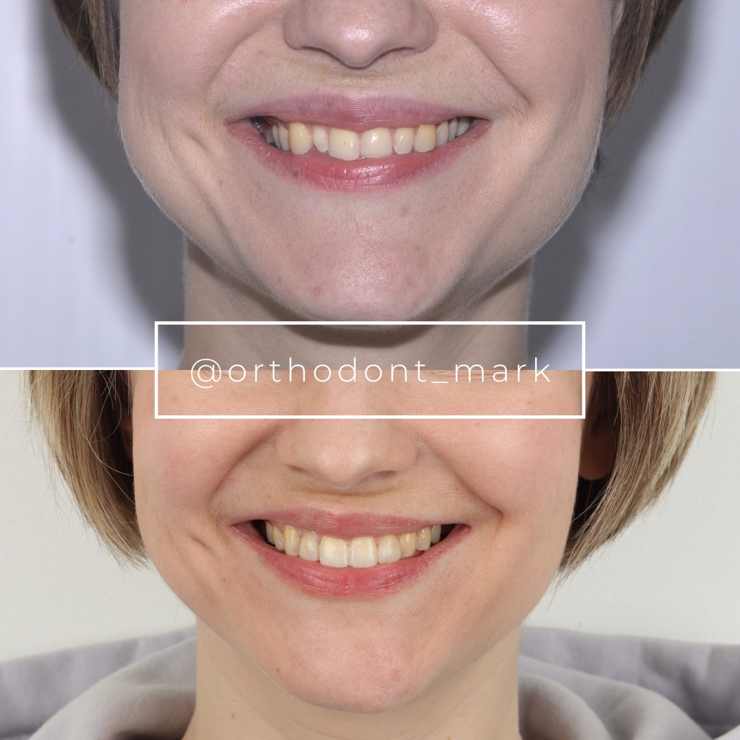
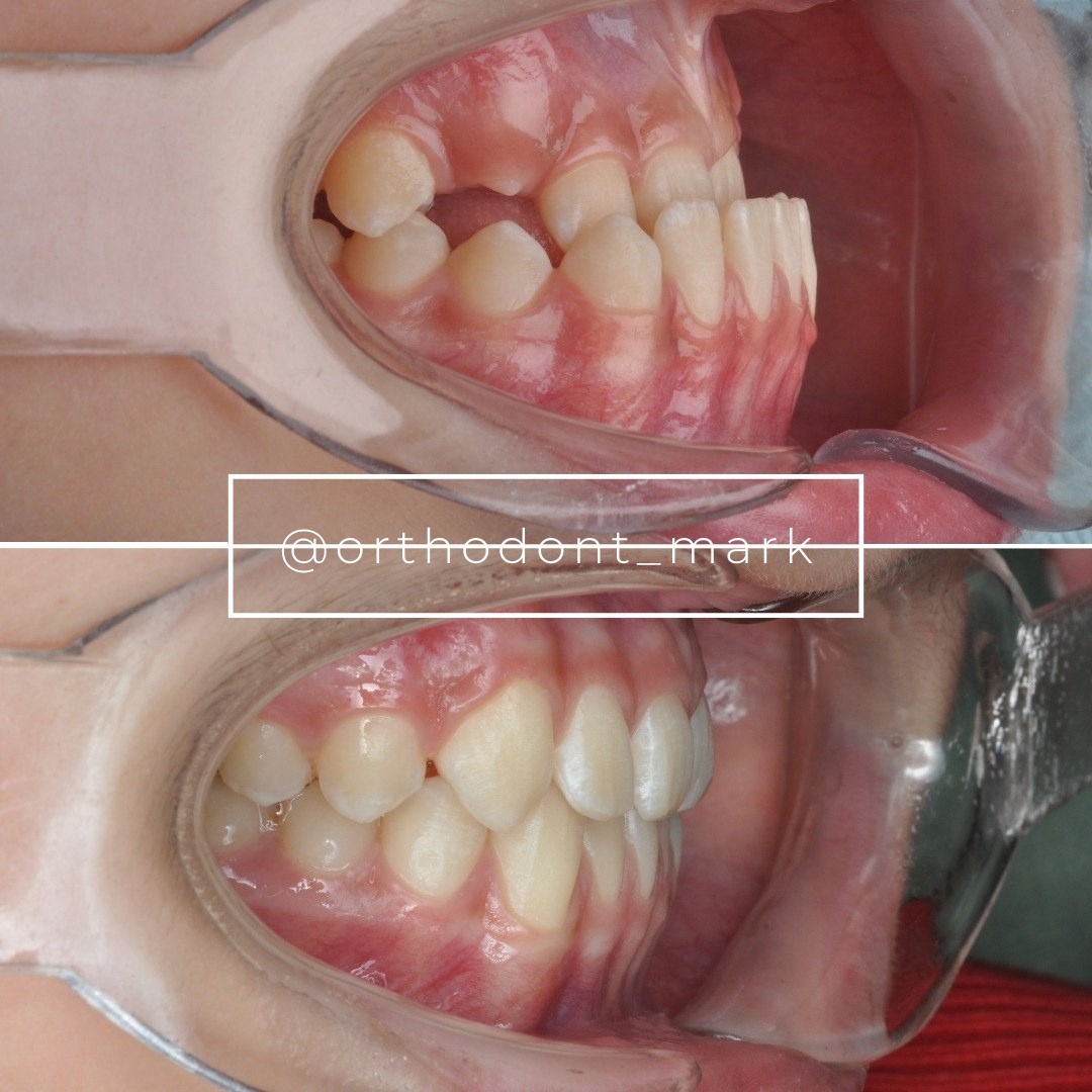

Learn the full MARPE protocol from Dr. Mark Radzhabov
Fundamental course covering CBCT patient selection, miniscrew planning, activation protocols, and 60+ clinical cases. Choose the access level that fits your practice.
Essentials of rapid palatal expansion for practicing orthodontists.
- Core RPE concepts and biomechanics
- 6 structured video lessons
- Clinical decision checklists
- Lifetime access to recordings
Deep-dive into MARPE protocol, diagnostics, and clinical execution.
- 6 modules covering MARPE from A to Z
- CBCT case selection walkthroughs
- Miniscrew placement and activation
- 60+ annotated clinical cases
- Direct Q&A with Dr. Mark Radzhabov
5-element medical consultation framework for dentists and orthodontists.
- Trust-building consultation protocol
- 5 lesson modules
- Templates for treatment plan delivery
- Works with any clinical specialty
Clinical FAQ
When to refer MARPE case review: what are the non-negotiable imaging triggers?
A non-negotiable referral trigger is a midpalatal suture staged D or E on CBCT, particularly when combined with concurrent pterygomaxillary or transpalatal fusion. Govaerts et al. (2023) documented that by age 15, 61% of females already present with such closed suture patterns, making CBCT-based staging the essential first step.
What referral thresholds distinguish a MARPE-eligible case from a surgical RPE case?
Expert consensus reported by Govaerts et al. (2023) placed surgical RPE cutoff ages at 15.1 years for orthodontists and 14.8 years for maxillofacial surgeons. Cases presenting at or above these thresholds with stage D or E suture maturation should be routed to surgical consultation before any bone-borne expansion is activated.
How does multi-provider orthodontic handoff documentation differ for MARPE versus conventional RPE?
MARPE handoffs require explicit documentation of miniscrew placement responsibility, regional suture staging, and activation management scope—items irrelevant in tooth-borne RPE. Chun et al. (2022) confirmed that MARPE produces significantly greater skeletal effects (p < 0.05), so the provider receiving the case must have the imaging baseline to manage those effects safely.
Which adjacent sutures should be staged on CBCT before initiating a MARPE referral?
Both the pterygomaxillary and transpalatal sutures should be staged alongside the midpalatal suture. Govaerts et al. (2023) reported pterygomaxillary closure of 83–100% and transpalatal closure of 78–85% by age 15 in females, meaning multi-suture fusion is common near the standard adolescent treatment window.
What activation data must accompany a MARPE case transferred mid-treatment?
The referring provider must document total turn count, weekly activation rate, and any asymmetric sutural response visible on sequential CBCT. Chun et al. (2022) used a standardized 35-turn expansion protocol with CBCT at baseline and post-expansion. Matching that documentation standard ensures the receiving specialist can assess the case without re-baselining.
How should a referring orthodontist verify skeletal outcomes after the case returns from a MARPE specialist?
Post-expansion CBCT at the 3-month consolidation point, per Chun et al. (2022), is the minimum standard. Comparing molar nasal width and palatine foramen dimensions against the specialist's immediate post-expansion record confirms whether skeletal separation was maintained before the referring provider assumes retention responsibility.
How do palatal expansion case complexity triage criteria change for female patients under 17?
Govaerts et al. (2023) found that pterygomaxillary and transpalatal suture closure accelerates rapidly between ages 13 and 15 in females, with midpalatal fusion at stages D and E in 61% by age 15. Triage criteria should be applied no later than early adolescence. Delayed CBCT assessment risks missing the non-surgical expansion window entirely.
What is the scope boundary between a referring orthodontist and a MARPE specialist in a multi-provider network?
The referring provider's scope covers CBCT acquisition, suture staging, transverse discrepancy measurement, and surgical triage flagging. The specialist's scope covers miniscrew placement planning, activation protocol management, and surgical escalation decisions. Documenting this boundary before treatment begins is what makes the handoff defensible.
Does suture maturation staging in younger patients require the same CBCT resolution as in adolescents near the surgical threshold?
Regional suture opening patterns are non-uniform even in younger patients. Kinzinger et al. (2022) examined CBCT and dental cast data in patients aged 7.3 to 13.8 years and found variable median palatal suture responses during RME, reinforcing that high-resolution imaging of anterior, middle, and posterior thirds is necessary regardless of patient age.
When should a second opinion on palatal expansion be sought before finalizing a MARPE treatment plan?
A second opinion is warranted whenever suture staging approaches D or E, the patient is near the expert-recommended cutoff ages of 15.1 years (orthodontist threshold) or 14.8 years (surgeon threshold) per Govaerts et al. (2023), or when the referring provider has not independently confirmed adjacent suture status on a current high-resolution CBCT.
Structured multi-provider handoff before MARPE is not a sign of limited scope. It is the evidence-based standard that peer-reviewed imaging data now demands. Suture fusion timelines documented by Govaerts et al. (2023) confirm that deferring case review until treatment stalls is too late—the triage window is narrow and CBCT-dependent. Orthodontists ready to build a defensible referral protocol will find structured frameworks and case-selection tools through Dr. Mark Radzhabov's resources at ortodontmark.com. Submitting borderline cases for specialist review before expansion activation is the single most protective step a clinician can take. Key sources: Chun et al. (2022), BMC Oral Health, doi:10.1186/s12903–022-02138-w. Govaerts et al. (2023), Journal of Orofacial Orthopedics, doi:10.1007/s00056–023-00487-x. Kinzinger et al. (2022), doi:10.1007/s00056–022-00429-z.