
Frontal Process Maxilla Resistance
Circummaxillary
Why Superior Anatomy Limits Skeletal Expansion
A clinician's anatomical guide to the resistance sites above the palate that determine MARPE and MSE outcomes.
TL;DR Frontal process maxilla resistance is a clinically underestimated barrier to MARPE skeletal expansion. The superior circummaxillary anatomy—including the nasomaxillary strut and frontonasal suture—dissipates lateral force before full sutural separation is achieved. Chun et al. (2022) confirmed suture separation in 95% of MARPE patients, yet skeletal gain varies with resistance anatomy above the palate.
When a MARPE appliance fails to produce expected skeletal widening despite confirmed midpalatal activation, the answer rarely lies in the palate itself. The frontal process maxilla resistance—transmitted superiorly through the nasomaxillary strut and circummaxillary suture network—constrains how much of the applied force actually translates into transverse skeletal displacement. At ortodontmark.com, Dr. Mark Radzhabov examines these superior resistance zones in clinical detail because case selection and force planning both depend on understanding anatomy that sits well above the miniscrew insertion plane. This article gives practicing orthodontists a framework for mapping those constraints before the first activation turn.
Which Circummaxillary Structures Actually Resist
Lateral
Force During Palatal Expansion?
The frontal process of the maxilla is the ascending bony pillar that articulates superiorly with the nasal bone and frontal bone, forming a rigid nasomaxillary strut that intercepts and dissipates lateral expansion forces before they can fully displace the maxillary halves. When a palatal expander is activated, the force vector is not confined to the midpalatal suture. It travels superiorly along the maxillary buttresses and encounters the circummaxillary suture network—frontonasal, nasomaxillary, and zygomaticomaxillary articulations—each adding cumulative resistance. Clinicians who map only the palatal suture on CBCT and ignore the superior resistance architecture risk misreading why expansion stalls or why skeletal gain is disproportionately small relative to appliance activation. The nasomaxillary strut is particularly consequential because its cortical thickness varies substantially across patients, independent of age. A dense strut in a younger adult can resist expansion more effectively than a thinner strut in an older patient, meaning that bone architecture above the insertion plane is a critical case-selection variable. Circummaxillary resistance zones collectively determine how much of the palatal force reaches the midpalatal suture as a true separating vector versus being absorbed in bending and micro-displacement of the surrounding buttresses. This distinction has direct implications for force magnitude planning in both MARPE and MSE protocols. The zygomaticomaxillary buttress adds a posterolateral component to the resistance pattern, while the frontonasal suture provides the most superior constraint. In patients with a high palatal vault and a pronounced frontal process, the effective moment arm between the force application point and the most resistant suture is longer, amplifying resistance torque. Appreciating this three-dimensional resistance geometry is foundational to the miniscrew-assisted skeletal expansion protocol and to understanding why equivalent activation turns can produce markedly different skeletal outcomes across patients.
What Do Separation Rates Tell Us About
Resistance
Above the Palate?
High midpalatal suture separation rates confirm that the appliance opened the palate—they do not confirm that superior resistance was overcome efficiently. Chun et al. (2022) reported midpalatal suture separation in 95% of MARPE patients and 90% of conventional RPE patients, with identical expansion amounts across groups. The near-equivalent separation rates are notable: a bone-borne appliance with miniscrews engaging cortical palatal bone still produced only a marginally higher separation frequency than a tooth-borne device, suggesting that the limiting variable is not anchor quality alone but the shared superior resistance anatomy both appliances must overcome. Chun et al. (2022) also found that MARPE produced significantly greater nasal width increases at the molar region and at the greater palatine foramen compared to conventional RPE, at a significance threshold of p < 0.05. This dimensional advantage implies that MARPE force delivery—because it bypasses the periodontal ligament and applies load closer to the basal bone—engages the circummaxillary resistance chain more directly, producing measurable deformation at nasal-level landmarks even when the superior buttresses are not surgically released. The practical implication is that a clinician choosing between appliance types should consider not only palatal suture response but also which superior anatomical sites the chosen force vector is most likely to engage. Sant'Ana et al. (2016) required a skeletal transverse maxillary deficiency exceeding 5 mm as an inclusion threshold, underscoring that clinically significant discrepancies—not borderline cases—are the target population for any expansion protocol that must overcome meaningful circummaxillary resistance. A marginal deficiency in a patient with dense frontal process cortex may respond adequately to conventional RPE, while a larger deficiency in the same anatomy might demand the superior force distribution that bone-borne MARPE or MSE provides.
Does SARME Override Frontal Process
Resistance
More Reliably Than MARPE Alone?
Surgically assisted rapid maxillary expansion directly addresses the superior resistance chain by osteotomizing the lateral maxillary walls, releasing the pterygomaxillary junction, and—when performed with midpalatal separation—sectioning the primary resistance structure at the palate. Sant'Ana et al. (2016) compared SARME with midpalatal split in 14 patients against SARME without midpalatal separation in 10 patients, providing direct evidence that the surgical protocol's completeness matters. Releasing only the lateral buttresses while leaving the midpalatal component intact alters force distribution through the remaining circummaxillary architecture, including the frontal process and frontonasal suture. The frontal process of the maxilla remains a passive resistance element even after lateral osteotomies unless the nasomaxillary strut is specifically addressed. This is why some surgeons extend the Le Fort I cut anteriorly to include the piriform rim region—targeting the base of the frontal process to reduce its buttressing effect. In purely MARPE-based protocols, this superior resistance zone is never surgically released, meaning that the appliance must generate sufficient force to produce micro-displacement through an intact nasomaxillary strut. Understanding this mechanical reality helps explain case-to-case variability in nasal width gain even when midpalatal separation is confirmed. For clinicians weighing conventional rapid palatal expander mechanics against bone-borne alternatives, the decision point is not merely suture maturity but the cumulative stiffness of the entire circummaxillary resistance chain. A patient with a high, thick frontal process and dense zygomaticomaxillary buttresses may achieve midpalatal separation with MARPE yet show limited nasal and transverse skeletal gain—an outcome that warrants early reassessment of the protocol rather than additional activation turns without imaging review.
How Does Appliance Anchor Type Alter
Force
Transmission Through the Resistance Chain?
The distinction between tooth-borne and bone-borne expansion lies not only in where anchorage is lost but in where along the circummaxillary resistance chain the applied force is introduced. A tooth-borne expander transmits force first through the periodontal ligament and alveolar process before any load reaches the basal maxillary bone and its superior buttresses. This compliance at the dental attachment point attenuates the effective skeletal component of the force vector—energy is consumed in tipping the maxillary premolars and molars outward rather than translating the basal bone laterally. Chun et al. (2022) confirmed that MARPE achieved significantly greater maxillary dental arch widths at both premolar and molar regions compared to conventional RPE (p < 0.05), yet the more important finding for the present discussion is that MARPE's bone-borne anchor introduces force directly at the palatal cortex, shifting the mechanical engagement point superiorly relative to the tooth roots. This means the frontal process resistance zone and the frontonasal suture are recruited earlier in the force chain, receiving a larger fraction of the total applied load than they would under tooth-borne expansion. The clinical corollary is that MARPE does not eliminate superior resistance—it engages it more directly. For MSE protocols using four palatal miniscrews positioned in the anterior and posterior palate, the insertion geometry further influences how force is distributed across the circummaxillary architecture. Anterior miniscrews engage a zone of bone that is structurally continuous with the base of the frontal process, so insertion depth and angulation relative to the palatal plane both influence how much force the nasomaxillary strut must absorb. Clinicians adding MARPE to their practice benefit from understanding this three-dimensional force path before selecting miniscrew positions on a case-by-case basis.
Integrating Superior Resistance Assessment Into
Pre-Treatment
Planning for MARPE and MSE
Pre-treatment CBCT evaluation should extend beyond midpalatal suture staging to include a systematic assessment of the superior resistance sites. The frontal process of the maxilla should be evaluated in the coronal plane for cortical thickness at the level of the inferior nasal aperture—this cross-section reliably represents the densest portion of the nasomaxillary strut. The frontonasal suture should be assessed for fusion status, since a fully obliterated suture converts an articulation into a continuous cortical barrier that no palatal appliance can deform without surgical assistance. When the CBCT reveals dense, fused superior circummaxillary anatomy alongside a partially mature midpalatal suture, the clinician faces a compound resistance problem: even if the palatal suture is mechanically separable, the superior chain may prevent adequate transverse skeletal displacement. In these cases, an intermediate protocol—such as MARPE with periodic CBCT reassessment at six to eight weeks—allows objective measurement of nasal width and intermolar skeletal distance before committing to continued activation or escalating to a surgical referral. Imaging-driven decision points are more reliable than activation count alone. Sant'Ana et al. (2016) set a transverse deficiency threshold of more than 5 mm for entry into their surgical expansion trial, a criterion that implicitly acknowledges the need for meaningful skeletal discrepancy to justify overcoming circummaxillary resistance through surgical means. Clinicians should apply an analogous logic to non-surgical MARPE planning: the greater the deficiency and the denser the superior resistance anatomy, the more critical it is to document skeletal—not just dental—response at an early timepoint and adjust the protocol accordingly.
Free Clinical Guide
Which Expansion Appliance at Which Age?
Get Dr. Mark Radzhabov's guide to skeletal-expansion appliances — indications, pros & cons, and how to choose by patient age. Free to your inbox.
Prefer video? Watch the free demo lesson on MARPE appliance types, features and trade-offs.
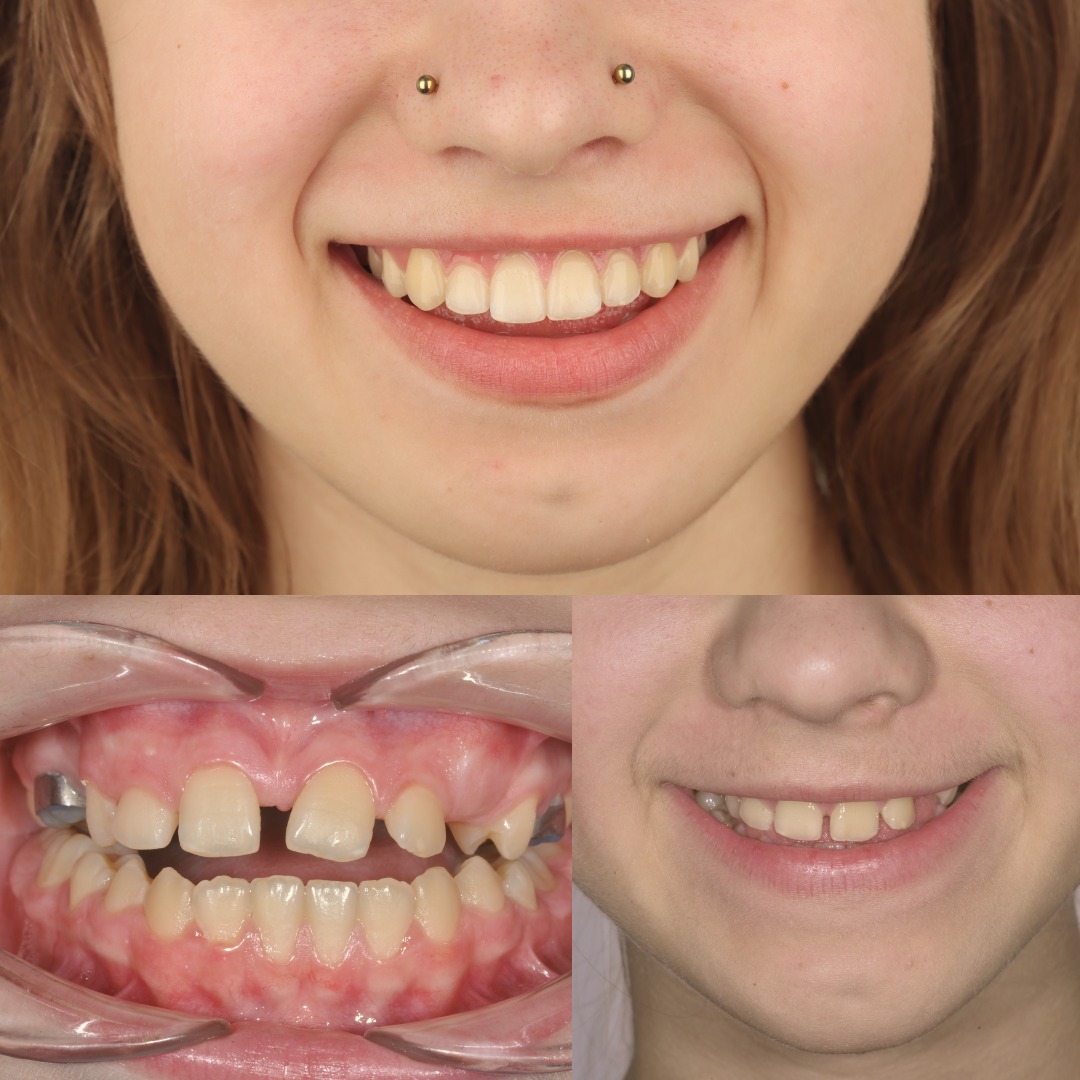
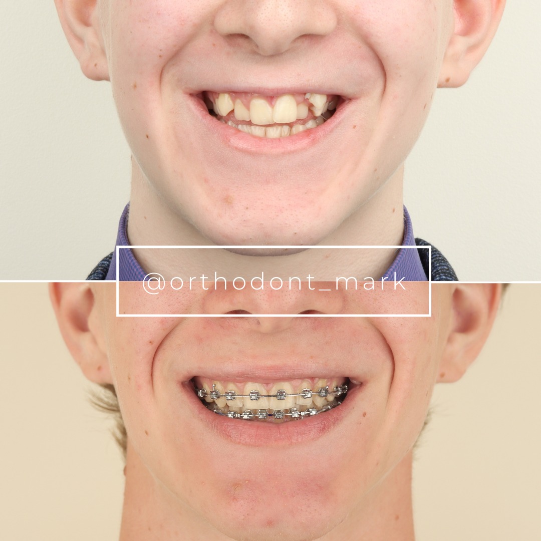
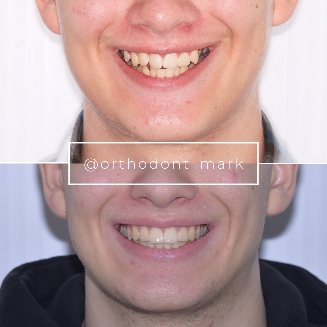
Documented outcomes
Clinical cases from Dr. Mark Radzhabov's practice
Learn the full MARPE protocol from Dr. Mark Radzhabov
Fundamental course covering CBCT patient selection, miniscrew planning, activation protocols, and 60+ clinical cases. Choose the access level that fits your practice.
Essentials of rapid palatal expansion for practicing orthodontists.
- Core RPE concepts and biomechanics
- 6 structured video lessons
- Clinical decision checklists
- Lifetime access to recordings
Deep-dive into MARPE protocol, diagnostics, and clinical execution.
- 6 modules covering MARPE from A to Z
- CBCT case selection walkthroughs
- Miniscrew placement and activation
- 60+ annotated clinical cases
- Direct Q&A with Dr. Mark Radzhabov
5-element medical consultation framework for dentists and orthodontists.
- Trust-building consultation protocol
- 5 lesson modules
- Templates for treatment plan delivery
- Works with any clinical specialty
Clinical FAQ
How does frontal process maxilla resistance affect MARPE skeletal expansion outcomes?
The frontal process forms a rigid nasomaxillary strut that absorbs lateral force before it fully separates the maxillary halves. Dense or fused frontal process cortex limits transverse skeletal displacement even when midpalatal suture separation is confirmed, making pre-treatment CBCT assessment of this structure essential for outcome prediction.
What circummaxillary suture anatomy should be evaluated before placing MARPE miniscrews?
Clinicians should assess the frontonasal, nasomaxillary, and zygomaticomaxillary sutures alongside the midpalatal suture. The frontonasal suture fusion status and frontal process cortical thickness are the most consequential superior variables because they determine how much force reaches the midpalatal suture as a true separating vector.
Does MARPE overcome nasomaxillary strut resistance better than conventional RPE?
Bone-borne MARPE engages the circummaxillary resistance chain more directly than tooth-borne RPE because force bypasses the periodontal ligament. Chun et al. (2022) confirmed greater nasal width gain with MARPE, indicating superior force transmission—but the frontal process resistance zone is still present and not surgically released.
At what minimum transverse deficiency does surgical expansion become justified over MARPE?
Sant'Ana et al. (2016) required a skeletal transverse maxillary deficiency exceeding 5 mm for inclusion in their surgical expansion trial. Deficiencies below this threshold in patients with moderate circummaxillary resistance may still respond to MARPE, but dense superior buttress anatomy shifts the threshold toward surgical assistance.
How does the frontonasal suture fusion status change the MARPE expansion force requirement?
A fully fused frontonasal suture converts the most superior articulation in the circummaxillary chain from a compliant joint into a rigid cortical barrier. This substantially increases the total resistance the palatal appliance must overcome, often producing poor skeletal-to-dental expansion ratios and warranting early reassessment of the non-surgical protocol.
Why does MARPE produce greater nasal width changes than RPE despite similar suture separation rates?
Bone-borne miniscrews deliver force directly to palatal cortex, engaging the basal maxilla and its nasal-level attachments more efficiently. Chun et al. (2022) found significantly greater nasal width at the molar region and greater palatine foramen with MARPE versus RPE (p < 0.05), reflecting more complete engagement of the superior force transmission pathway.
How should CBCT imaging criteria guide the decision between MARPE and SARME in adults?
CBCT should assess midpalatal suture maturity, frontal process cortical density, and frontonasal suture fusion. When the superior circummaxillary anatomy shows dense, fused buttresses alongside an advanced-stage midpalatal suture, SARME provides direct surgical release of the lateral and superior resistance chain that MARPE cannot overcome non-surgically.
What is the clinical significance of greater palatine foramen width change as a MARPE outcome measure?
The greater palatine foramen sits at the posterior palatal base and its widening reflects true basal bone displacement rather than dental or alveolar tipping. Chun et al. (2022) identified significantly greater foramen width change with MARPE versus RPE, confirming that bone-borne force engages the skeletal resistance architecture more effectively.
How does anterior miniscrew placement in MSE interact with the frontal process resistance zone?
Anterior palatal miniscrews engage bone structurally continuous with the base of the frontal process. Their insertion depth and angulation relative to the palatal plane influence how much of the applied force is transmitted superiorly through the nasomaxillary strut versus directed laterally toward the midpalatal suture, making anterior miniscrew geometry a critical biomechanical variable.
When is mid-treatment CBCT reassessment warranted during MARPE in patients with high circummaxillary resistance?
When pre-treatment imaging reveals dense frontal process cortex or fused superior sutures, reassessment at six to eight weeks allows objective measurement of nasal width and skeletal intermolar distance. Absence of measurable skeletal response by that checkpoint—rather than activation count—is the most reliable indication to escalate toward surgical consultation.
Understanding how the frontal process of the maxilla and the broader circummaxillary suture network limit palatal expansion is not academic—it directly shapes whether a given patient needs MARPE, a heavier-force MSE protocol, or surgical assistance. Suture maturation, bone density, and superior buttress anatomy must be integrated into every pre-treatment CBCT review. Dr. Mark Radzhabov invites clinicians to explore structured MARPE training at ortodontmark.com, submit a case for review, or consult directly on challenging expansion candidates. Sound anatomical reasoning before appliance placement is the single most reliable predictor of a stable skeletal outcome. Key sources: Sant'Ana et al. (2016), Int J Oral Maxillofac Surg, doi:10.1016/j.ijom.2016.03.005. Chun et al. (2022), BMC Oral Health, doi:10.1186/s12903–022-02138-w.