
Palatal Bone Volume at the Third Rugae Region
Anatomic Evidence for Miniscrew Placement
Evidence-based imaging criteria and site-selection logic for reliable palatal miniscrew anchorage in MARPE protocols.
TL;DR Palatal bone volume at the third rugae region is a critical anatomic determinant for miniscrew placement in MARPE. Bone density and cortical thickness at this landmark directly influence primary stability and suture-separation outcomes. Kapetanovi et al. (2021) reported an overall MARPE success rate of 92.5%, with skeletal width gains of 2.33 mm—outcomes that hinge on sound screw positioning.
Selecting the precise insertion site for palatal miniscrews remains one of the most consequential steps in planning a MARPE case, yet it rarely receives the anatomic granularity it deserves. The third rugae region has emerged as a reliable surface landmark for guiding anterior palatal screw placement, correlating with a zone of adequate cortical bone volume away from the nasopalatine canal and root apices. In this article, Dr. Mark Radzhabov of ortodontmark.com reviews the anatomic evidence, imaging criteria, and site-selection logic that underpin safe and stable miniscrew anchorage at this location.
Why Does the Third Rugae Region Matter for
Miniscrew Placement?
The third rugae region is an intraoral surface landmark on the anterior palate, located at the level of the third transverse palatal rugae, that demarcates a zone of comparatively dense cortical bone suitable for primary miniscrew anchorage in bone-borne expansion appliances. This position sits anterior to the greater palatine foramen, lateral to the nasopalatine canal, and coronal to the root apices of the premolars—three anatomic boundaries that define the safe zone for screw insertion without violating neurovascular structures or compromising periodontal support. Cone-beam CT cross-sections through this landmark reveal that the palatal vault at the third rugae level typically displays the greatest buccopalatal cortical thickness available in the anterior-to-middle palate corridor. Thicker cortex translates directly into higher insertion torque values, reduced micromotion at the bone–implant interface, and more reliable primary stability before osseointegration—each of which is a prerequisite for sustaining the activation forces used in MARPE appliance design and skeletal expansion biomechanics. The practical implication is straightforward: placing screws too far posteriorly risks engaging the thinner cancellous bone overlying the middle palatal suture, while placement too far anteriorly encroaches on the incisive foramen. The third rugae landmark provides a reproducible, visually confirmable reference that any clinician can identify chairside and then verify on pre-treatment CBCT before finalizing screw coordinates.
How Does Placement Site Influence Suture Separation
and Skeletal Gain?
The quality of miniscrew anchorage at the chosen palatal site determines how cleanly activation force is transmitted across the midpalatal suture rather than dissipated through dental tipping or screw micromotion. Kapetanovi et al. (2021) reported a pooled MARPE success rate of 92.5% (95% CI: 88.7–96.3%), a mean skeletal width increase of 2.33 mm (95% CI: 1.63–3.03 mm), and a dental intermolar width increase of 6.55 mm (95% CI: 5.50–7.59 mm)—figures that reflect protocols in which bone-borne force was the primary mechanical pathway. Sex and skeletal maturity modulate those aggregate numbers considerably. Jeon et al. (2022) found that suture separation occurred in 94.17% of female patients versus 61.05% of males, with an overall rate of 79.53% across their 215-patient sample. Critically, older age correlated significantly with suture non-separation in males (p = 0.001) but not in females (p = 0.221), indicating that the force pathway established by screw positioning interacts with patient-level biological variables rather than acting as the sole determinant. Stable anterior palatal anchorage does not override maturation status—it maximizes the probability of separation in patients whose suture biology is amenable. Chun et al. (2022) confirmed that MARPE produced significantly less buccal displacement of premolar and molar anchor teeth than conventional RPE, a finding that is mechanistically inseparable from the bone-borne force distribution enabled by well-positioned palatal screws. When screws lack primary stability—whether from inadequate bone volume or suboptimal site selection—the appliance shifts load toward the dental anchor bands, reintroducing the dentoalveolar tipping that bone-borne design is specifically engineered to eliminate.
What Imaging Criteria Confirm the Third Rugae Site
Before Insertion?
Pre-treatment CBCT is the diagnostic standard for evaluating palatal bone volume at the third rugae region. A cross-sectional reconstruction in the coronal plane at the level of the third rugae allows direct measurement of cortical thickness on both the nasal and oral surfaces of the palatal shelf, identification of cancellous bone depth, and precise localization of the nasopalatine canal in three dimensions. The region-of-interest cursor should be centered at the intersection of the palatal midline and the transverse plane through the rugae to standardize measurements across sequential imaging. Hounsfield unit values extracted from this ROI give a density estimate that correlates with expected insertion torque and primary stability. Thin cancellous bone with low HU values at the intended site is a signal to shift the planned insertion point anteriorly—where the cortex is typically denser—or to counsel the clinician toward a tooth-borne or hybrid appliance design. Comparing left and right palatal shelves at the same axial level also reveals asymmetric bone volume, which can inform differential screw angulation to avoid root proximity on the thinner side. Kapetanovi et al. (2021) noted that MARPE expansion duration ranged from 20 to 126 days across the included studies, a span that reflects substantial variation in activation protocols and patient response. Imaging that confirms adequate bone volume at placement allows clinicians to sustain those longer activation windows with confidence that screws will remain stable rather than loosening under cumulative load. For a practical walkthrough of how imaging findings integrate with appliance selection, the rapid palatal expander tooth-borne force comparison resource provides complementary context.
Does Anterior Palatal Anchorage Change How Force
Reaches the Suture?
The mechanical rationale for targeting the third rugae region is rooted in force-vector geometry. Screws placed at this anterior position deliver activation forces along a trajectory that engages the anterior palatal suture preferentially, where the fibrous interdigitation is typically less calcified and more amenable to gradual separation than in the middle or posterior thirds. This anterior force concentration also positions the resultant vector closer to the midpalatal suture's centroid of resistance, reducing the moment arm that would otherwise generate asymmetric opening or suture rocking. Chun et al. (2022) demonstrated that MARPE produced greater increases in nasal width and palatine foramen dimensions compared to conventional RPE, a skeletal footprint that is consistent with force being distributed through palatal bone rather than through dental crowns and periodontium. The reduced buccal displacement of anchor teeth observed in the same study is the clinical corollary: when screws are stable and correctly positioned, the appliance behaves as designed—bone-borne rather than tooth-borne. Jeon et al. (2022) also confirmed that suture separation in their 215-patient sample varied markedly by sex, with male patients showing significantly lower separation rates (61.05%) than females (94.17%). While this sex difference involves biological variables beyond screw position alone, it reinforces the principle that maximizing every controllable mechanical advantage—including optimal anterior palatal anchorage—is essential when treating patient groups where suture biology is less favorable. Clinicians planning their first bone-borne cases can explore the complete decision framework through structured MARPE training and mentorship at ortodontmark.com.
How Does Patient Age and Sex Modify the Bone-Volume
Equation?
Screw-site selection does not exist in a vacuum. It interacts continuously with the patient's skeletal maturity, sex, and suture histology. Jeon et al. (2022) demonstrated that across their retrospective cohort of 215 patients ranging from 6 to 60 years of age—95 male and 120 female—older age was a significant predictor of suture non-separation in male patients (p = 0.001) but showed no significant association in female patients (p = 0.221). This asymmetry suggests that hormonal or structural differences in suture fusion tempo mean that equivalent bone-volume and screw-position quality will produce different mechanical outcomes depending on the patient's biological profile. For the clinician, this finding translates into a tiered risk assessment rather than a binary pass/fail. A male patient in whom CBCT reveals borderline cortical thickness at the third rugae site carries compounded risk: marginal bone volume threatens screw stability, while age-related suture calcification reduces the probability that even perfect force transmission will yield separation. In these cases, the imaging evidence should prompt earlier counseling about alternative approaches rather than proceeding optimistically. Conversely, Chun et al. (2022) showed that midpalatal suture separation occurred in 95% of MARPE patients versus 90% of RPE patients in their randomized trial, confirming that bone-borne protocols maintain a meaningful separation advantage when patient selection and screw positioning are both optimized. The aggregate data from Kapetanovi et al. (2021) further support this: a 92.5% pooled success rate is achievable when site selection, imaging assessment, and appliance design converge on an evidence-based protocol.
Free Clinical Guide
Which Expansion Appliance at Which Age?
Get Dr. Mark Radzhabov's guide to skeletal-expansion appliances — indications, pros & cons, and how to choose by patient age. Free to your inbox.
Prefer video? Watch the free demo lesson on MARPE appliance types, features and trade-offs.
Documented outcomes
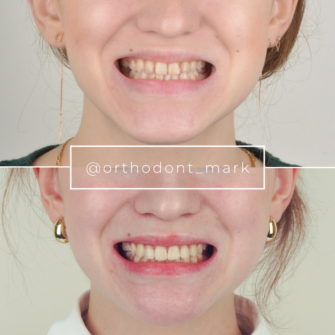
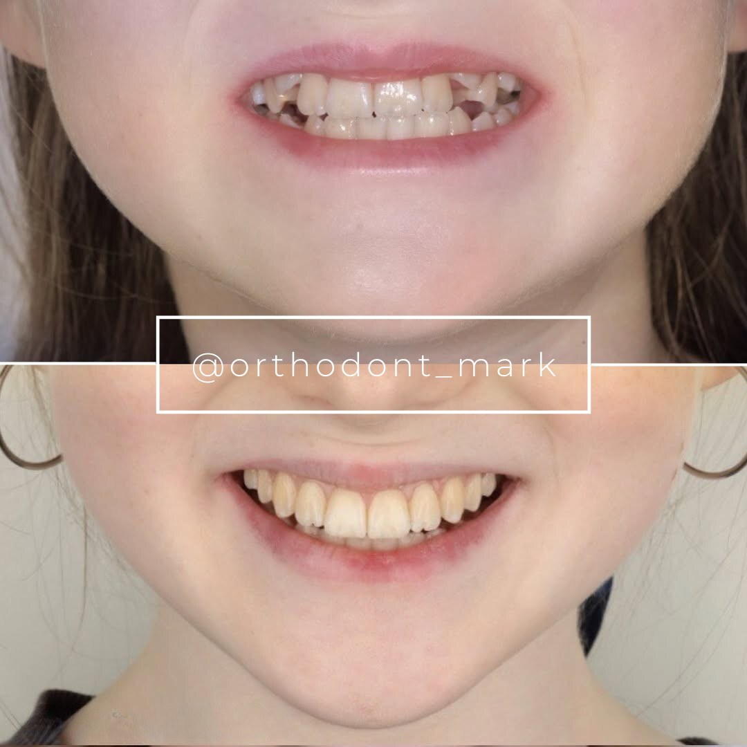
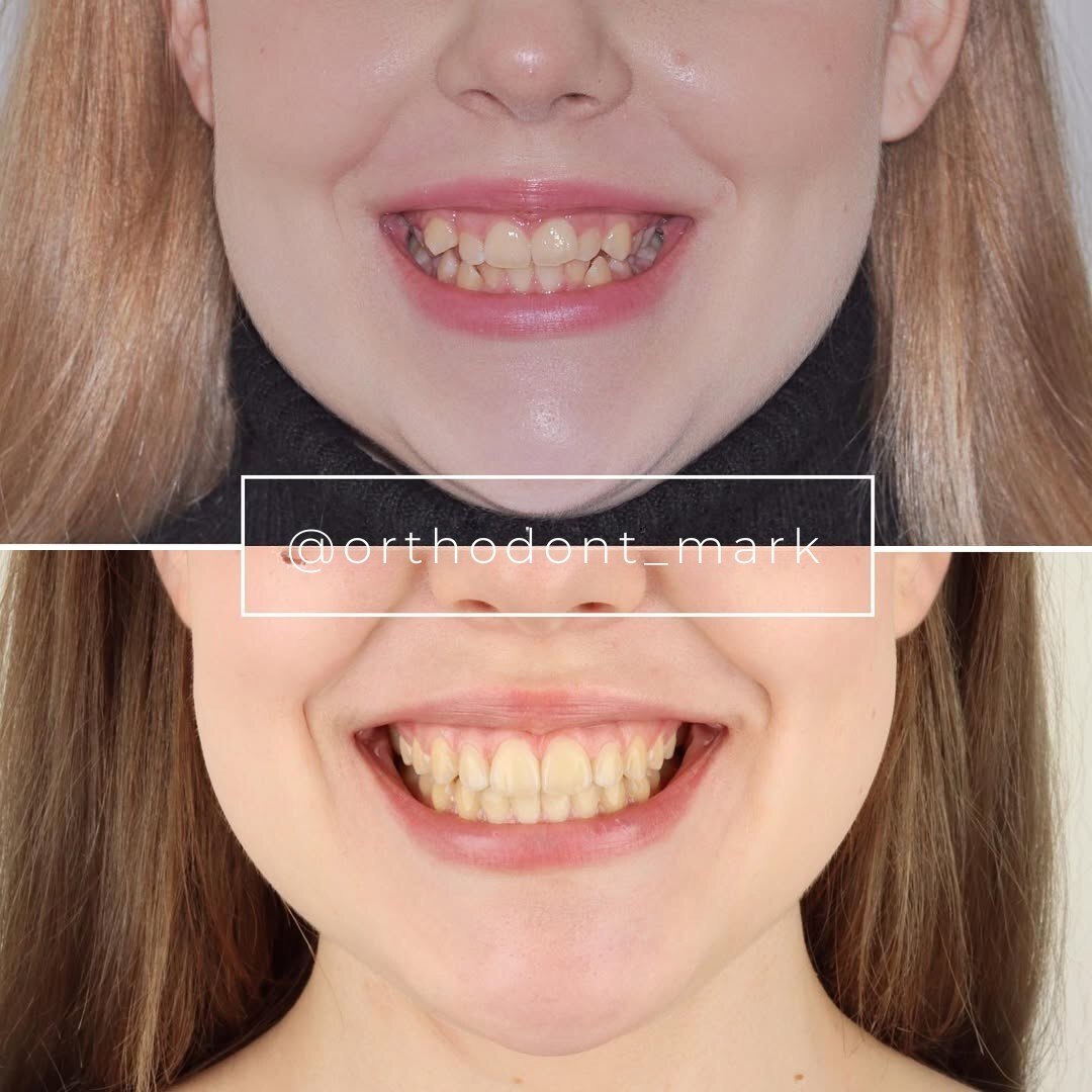
Clinical cases from Dr. Mark Radzhabov's practice
Learn the full MARPE protocol from Dr. Mark Radzhabov
Fundamental course covering CBCT patient selection, miniscrew planning, activation protocols, and 60+ clinical cases. Choose the access level that fits your practice.
Essentials of rapid palatal expansion for practicing orthodontists.
- Core RPE concepts and biomechanics
- 6 structured video lessons
- Clinical decision checklists
- Lifetime access to recordings
Deep-dive into MARPE protocol, diagnostics, and clinical execution.
- 6 modules covering MARPE from A to Z
- CBCT case selection walkthroughs
- Miniscrew placement and activation
- 60+ annotated clinical cases
- Direct Q&A with Dr. Mark Radzhabov
5-element medical consultation framework for dentists and orthodontists.
- Trust-building consultation protocol
- 5 lesson modules
- Templates for treatment plan delivery
- Works with any clinical specialty
Clinical FAQ
What is the third rugae region and why is it used as a landmark for palatal miniscrew placement?
The third rugae region marks the level of the third transverse palatal rugae in the anterior palate. It overlies a zone of comparatively dense cortical bone with adequate clearance from the nasopalatine canal and root apices, making it a reliable, visually confirmable reference for MARPE screw insertion.
How does palatal bone volume at the third rugae region affect primary miniscrew stability?
Greater cortical thickness at this site increases insertion torque and reduces micromotion at the bone–implant interface. Thin or low-density bone—identifiable by Hounsfield unit mapping on CBCT—predicts reduced primary stability and a higher risk of screw loosening under sustained activation forces.
What CBCT imaging criteria should I evaluate before placing MARPE screws at the anterior palate?
Assess cortical thickness and Hounsfield unit density in coronal cross-section at the third rugae level, confirm lateral clearance from the nasopalatine canal on axial slices, and verify vertical clearance from lateral incisor and canine apices on sagittal reconstructions before finalizing screw coordinates.
How does the MARPE suture separation success rate compare between male and female patients?
Jeon et al. (2022) reported suture separation in 94.17% of female patients versus 61.05% of male patients in a 215-patient sample. Older age significantly predicted non-separation in males (p = 0.001) but not females (p = 0.221), indicating sex-specific biological variation beyond screw position.
What overall MARPE success rate does the meta-analytic evidence support?
Kapetanovi et al. (2021) reported a pooled MARPE success rate of 92.5% (95% CI: 88.7–96.3%), with a mean skeletal width increase of 2.33 mm and dental intermolar width increase of 6.55 mm across the included studies.
How does MARPE compare to conventional RPE for suture separation and dental side effects?
Chun et al. (2022) found suture separation in 95% of MARPE patients versus 90% of RPE patients, while MARPE produced significantly less buccal displacement of premolar and molar anchor teeth. Bone-borne force transmission reduces dentoalveolar tipping compared to tooth-borne appliances.
How long does the MARPE expansion phase typically last according to published evidence?
Kapetanovi et al. (2021) noted that MARPE expansion duration ranged from 20 to 126 days across pooled studies, reflecting variation in activation protocols, appliance design, and patient response. Screw stability at the insertion site is critical for sustaining longer activation windows.
Does anterior palatal miniscrew placement reduce dental tipping compared to tooth-borne RPE?
Yes. Chun et al. (2022) confirmed that MARPE produced significantly less buccal displacement of premolar and molar anchor teeth than conventional RPE during expansion and consolidation, a direct mechanical consequence of distributing force through palatal bone rather than the periodontal ligament.
When should borderline palatal bone volume at the third rugae site prompt a change in treatment approach?
When CBCT reveals low Hounsfield unit density or thin cortex at the third rugae site in a male patient showing age-related suture changes, the compounded risk of screw failure and non-separation should prompt earlier discussion of surgical assistance or a tooth-borne hybrid design.
How does MARPE skeletal width gain differ from dental intermolar width change?
Kapetanovi et al. (2021) reported mean skeletal width increase of 2.33 mm and dental intermolar width increase of 6.55 mm, confirming that the majority of measured expansion is dentoalveolar. True skeletal gain requires confirmed midpalatal suture separation, underscoring the importance of bone-borne anchorage quality.
Accurate identification of palatal bone volume at the third rugae region is not a refinement—it is a prerequisite for predictable MARPE outcomes across the age spectrum documented in the literature. The convergence of suture-separation data from Jeon et al. (2022), Chun et al. (2022), and Kapetanovi et al. (2021) underscores how biomechanically sound screw positioning amplifies every other variable in the protocol. Dr. Mark Radzhabov invites colleagues to submit a case for review or enroll in structured mentorship at ortodontmark.com, where site-selection imaging and appliance sequencing are covered in clinical depth. Key sources: Jeon et al., 2022, Clinical Oral Investigations, doi:10.1007/s00784–021-04281–0. Kapetanovi et al., 2021, The European Journal of Orthodontics, doi:10.1093/ejo/cjab005. Chun et al., 2022, BMC Oral Health, doi:10.1186/s12903–022-02138-w.

