When to Refer a MARPE Case
Red Flags
That Signal Limits of In-Office Capability
A structured checklist of documented contraindications and imaging findings that tell you when expansion management must move beyond a solo in-office protocol.
TL;DR Recognizing when a MARPE case exceeds in-office capability requires systematic evaluation of suture morphology, facial trauma history, jaw pathology, and appliance biomechanics. Documented red flags—asymmetric suture, foreign bodies in the expansion field, cleft-related deformities—signal that referral for case review or surgical consultation is clinically warranted before treatment begins.
Not every transverse maxillary deficiency is an uncomplicated MARPE case, and the consequences of misidentifying a high-risk expansion patient compound quickly once the activation protocol is underway. At ortodontmark.com, Dr. Mark Radzhabov teaches orthodontists to audit each case against a structured set of contraindications and imaging criteria before placing a single TAD. This article distills those documented red flags—drawn from peer-reviewed evidence and Dr. Radzhabov's clinical framework—so practitioners can make a confident, early decision: proceed, modify, or refer for specialist case review.
What Makes a MARPE Case
Too Complex
to Manage In-Office?
A MARPE case that exceeds in-office capability is one in which documented anatomical, pathological, or biomechanical red flags raise the probability of asymmetric expansion, TAD failure, or surgical-level complications beyond what can be safely managed within a standard orthodontic workflow. The distinction matters because MARPE's bone-borne force delivery—bypassing anchor teeth—amplifies the skeletal consequences of any planning error. An unrecognized asymmetric suture, for example, does not merely produce a suboptimal result. It produces a midline shift and occlusal cant that requires multidisciplinary correction. Dr. Radzhabov's protocol identifies four primary contraindication categories that, when present, require either modified appliance design, surgical co-management, or formal referral for case review: suture asymmetry, facial trauma history, jaw structural pathology, and material allergy. These categories are not abstract—each has a specific clinical presentation, a specific imaging correlate on CBCT, and a specific downstream complication if overlooked. Suture maturation stage adds a fifth decision layer: Govaerts et al. (2023) found midpalatal suture closure at stages D and E in 61% of 15-year-old females, a finding that recasts age as an unreliable proxy and makes CBCT morphology the true triage criterion. Before any activation protocol begins, the treating orthodontist must confirm that none of these red flags are present. When they are, the appropriate response is documented escalation—not improvisation under load.
How Suture Closure Stage
Changes
the Referral Threshold
Chronological age is a weak triage filter for skeletal expansion planning. Govaerts et al. (2023) demonstrated that the pterygomaxillary suture shows 83–100% closure in females aged 13 to 17 years, meaning that even in early adolescence a substantial proportion of patients carry suture morphology that resists conventional expansion forces. The same study found that clinical observers recommended switching from orthopedic to surgically assisted expansion at cutoff ages of 15.1 years for orthodontists and 14.8 years for surgeons—a narrow window that underscores how quickly the indication for SARPE emerges. Chun et al. (2022) provide the skeletal expansion benchmark: midpalatal suture separation occurred in 95% of MARPE cases and 90% of RPE cases under study conditions. Those figures represent best-case outcomes in selected, protocol-appropriate patients—not the realistic population that includes unscreened suture stages. When CBCT reveals advanced suture closure, the probability of achieving comparable separation with a bone-borne appliance alone drops, and the risk of generating exclusively dentoalveolar displacement—with its attendant buccal bone dehiscence—rises markedly. The practical referral trigger is not a birthday. It is the CBCT finding of bilateral cortical bridging across the midpalatal suture in the posterior and middle thirds, combined with a clinical presentation that demands more than minimal skeletal expansion. Any such case warrants either a SARPE consultation or, at minimum, a multidisciplinary case review before the expansion screw is touched.
When Does Appliance Force Output
Signal
a Case Beyond In-Office Management?
Screw selection directly determines the force envelope delivered to the midpalatal suture, and that envelope must be matched to the patient's bone resistance—not defaulted to whatever device is available in the laboratory. Camporesi et al. (2013) measured in vitro force generation across expansion screw designs and found that Hyrax and A2620 screws generated forces exceeding 20 kg, while Palatal Split screws generated approximately 16 kg. A2620 and Hyrax screws also demonstrated significantly greater mechanical rigidity than Palatal Split screws across all activation levels (Camporesi et al., 2013). In a patient with advanced suture ossification, applying a 20-kg-range force without surgical cortical release risks TAD instability, anchor tooth trauma, or device failure rather than clean sutural opening. Dr. Radzhabov's protocol flags TAD loosening as a critical intraoperative sign that skeletal support has been lost. When TADs are inadequate—too short, improperly angulated, or placed in low-density cortical bone—the full expansion load redirects to anchor teeth, converting a planned skeletal correction into dentoalveolar tipping. Chun et al. (2022) confirmed that MARPE produces significantly less buccal displacement of anchor teeth compared to RPE (p<0.05), but that advantage evaporates when TAD stability is compromised. Any case in which preoperative CBCT reveals palatal bone thickness insufficient for the target TAD insertion depth, or in which intraoperative probing confirms inadequate cortical purchase, should trigger immediate escalation. Continuing to activate an unstable construct in hopes of a late skeletal response is a planning error—not a clinical option.
Jaw Pathology and Cleft Anatomy:
Absolute
vs. Relative Contraindications
Cleft lip and palate deformities, partial palatal defects, and significant asymmetric alveolar bone loss represent the structural pathology category in Dr. Radzhabov's contraindication framework. These cases share a common problem: the maxillary halves do not present equivalent bone volume or density for TAD anchorage, making symmetric force application geometrically impossible with a standard MARPE design. In some presentations, TAD placement itself is anatomically precluded by scar tissue or bone discontinuity at the palatal vault. Dr. Radzhabov acknowledges that structural jaw pathology is not an absolute contraindication in every instance—the final decision belongs to the examining clinician after full CBCT review. The operative principle, however, is that any case where standard four-TAD MARPE geometry cannot be replicated symmetrically on both sides of the suture requires a formal multidisciplinary assessment. Attempting expansion with asymmetric TAD distribution predictably produces differential force vectors, unequal sutural opening, and midline deviation. The referral trigger for this category is straightforward: if the preoperative CBCT reveals that planned TAD insertion sites on one side of the midpalatal suture are within scar tissue, adjacent to a cleft margin, or overlying a bone graft, the case requires input from an oral and maxillofacial surgeon before any appliance is fabricated. Fabricating the device first and discovering the anatomical problem at the insertion appointment wastes clinical time and erodes patient confidence.
Building a Referral-Ready
Documentation
Protocol for High-Risk Expansion Cases
A referral for case review carries clinical weight only when it is accompanied by structured documentation: a full CBCT with suture staging noted, a photograph protocol baseline, a written contraindication checklist, and a specific clinical question for the receiving specialist. Sending a patient to a maxillofacial surgeon with a vague concern about suture density is not a referral—it is a handoff without a diagnostic anchor. The CBCT request should specify midpalatal suture morphology in anterior, middle, and posterior thirds. TAD insertion site bone depth at the planned coordinates. And the presence or absence of prior hardware or osseous pathology. For cases approaching the pterygomaxillary suture closure window that Govaerts et al. (2023) identified—with orthodontists recommending a shift to surgically assisted expansion at 15.1 years—the referral should be timed before the suture stages advance further, not after a failed expansion attempt. Waiting for clinical signs of failure (TAD loosening, asymmetric diastema, occlusal cant development) converts a preventable problem into a corrective one. Dr. Radzhabov's protocol also includes patient communication as a documentation element: patients should be informed of the anatomical findings, the reason for referral, and the treatment pathway that follows. This is not only an ethical requirement—it sustains case continuity when the patient returns after a surgical consultation. Orthodontists who build a referral-ready workflow lose fewer cases to attrition and retain clinical ownership of the expansion phase even when surgical assistance is required.
Free Clinical Guide
Which Expansion Appliance at Which Age?
Get Dr. Mark Radzhabov's guide to skeletal-expansion appliances — indications, pros & cons, and how to choose by patient age. Free to your inbox.
Prefer video? Watch the free demo lesson on MARPE appliance types, features and trade-offs.
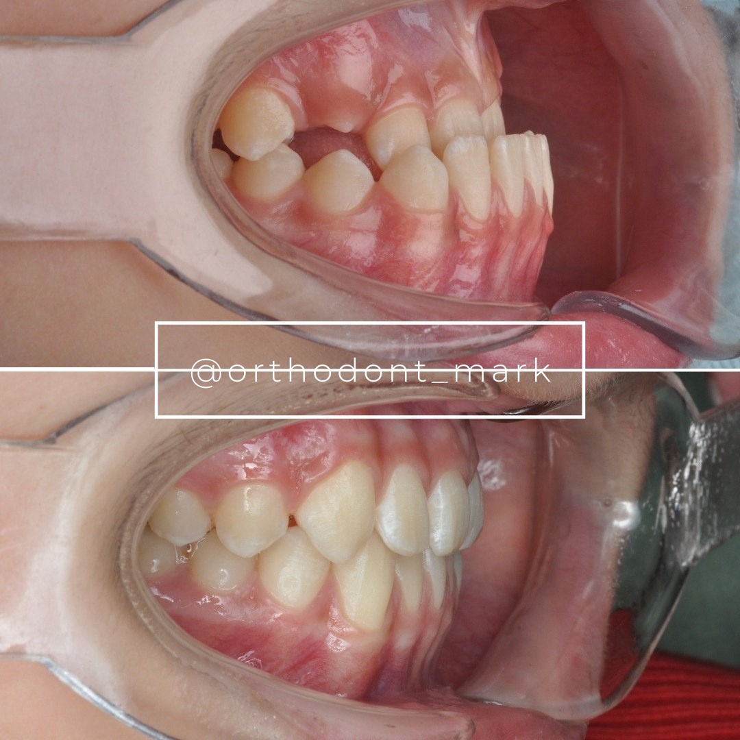
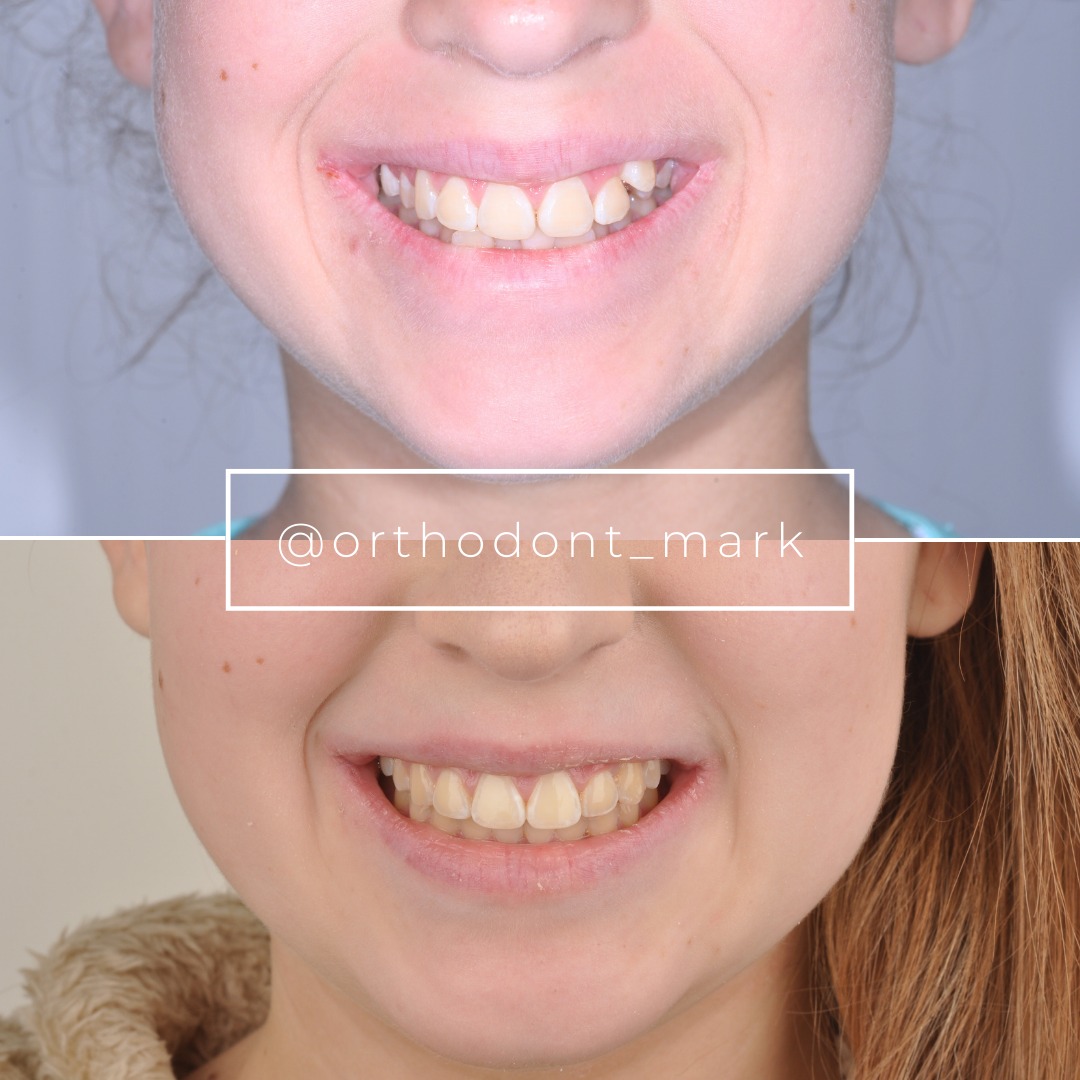
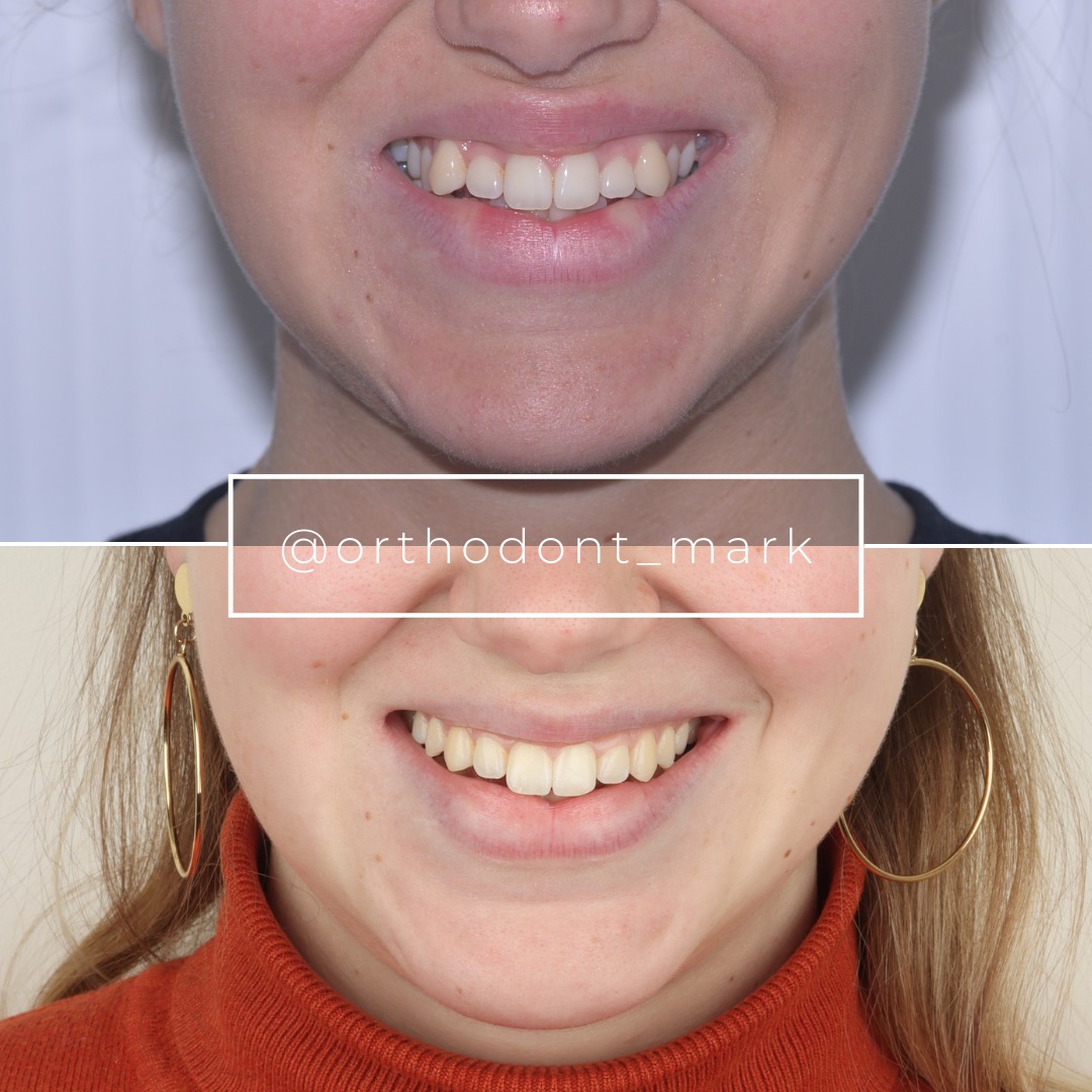
Documented outcomes
Clinical cases from Dr. Mark Radzhabov's practice
Learn the full MARPE protocol from Dr. Mark Radzhabov
Fundamental course covering CBCT patient selection, miniscrew planning, activation protocols, and 60+ clinical cases. Choose the access level that fits your practice.
Essentials of rapid palatal expansion for practicing orthodontists.
- Core RPE concepts and biomechanics
- 6 structured video lessons
- Clinical decision checklists
- Lifetime access to recordings
Deep-dive into MARPE protocol, diagnostics, and clinical execution.
- 6 modules covering MARPE from A to Z
- CBCT case selection walkthroughs
- Miniscrew placement and activation
- 60+ annotated clinical cases
- Direct Q&A with Dr. Mark Radzhabov
5-element medical consultation framework for dentists and orthodontists.
- Trust-building consultation protocol
- 5 lesson modules
- Templates for treatment plan delivery
- Works with any clinical specialty
Clinical FAQ
What are the absolute contraindications for MARPE that require specialist referral?
Per Dr. Radzhabov's protocol, documented contraindications include asymmetric midpalatal suture morphology, any prior facial trauma or retained maxillofacial hardware in the expansion field, jaw structural pathology such as cleft palate deformities, and confirmed allergy to appliance materials. Each warrants referral or surgical co-management before treatment begins.
How does CBCT suture staging change the decision to refer a MARPE case to surgery?
When CBCT reveals bilateral cortical bridging in the posterior and middle suture thirds, the probability of clean sutural opening with bone-borne force alone drops substantially. Govaerts et al. (2023) found that clinicians recommended shifting to surgically assisted expansion at 15.1 years (orthodontists) and 14.8 years (surgeons), based on observed suture closure patterns.
What intraoperative sign during MARPE activation indicates the case has exceeded in-office capability?
TAD mobility—identified indirectly by peri-implant mucosal inflammation on photo-protocol review—signals that skeletal anchorage has been lost. Without intact TAD stability, the expansion load shifts entirely to anchor teeth, producing dentoalveolar rather than skeletal displacement. Continued activation at that point is a planning error requiring immediate case review.
When should I refer a MARPE patient for SARPE evaluation based on suture maturation?
Refer before activation when CBCT shows advanced midpalatal suture ossification and the clinical expansion demand is substantial. Govaerts et al. (2023) document that midpalatal suture closure was present in 61% of 15-year-old females—making mid-adolescence a realistic SARPE referral window, not solely adulthood.
Does prior maxillofacial fracture always contraindicate MARPE?
Not categorically, but any retained hardware—plates, screws, or fracture callus—in the planned expansion field acts as a counterforce and may produce asymmetric sutural opening. Dr. Radzhabov cites a colleague's case where an undetected cheekbone fracture caused asymmetric expansion. Full imaging of prior trauma is mandatory before case acceptance.
How does expansion screw selection relate to referral criteria in high-resistance sutures?
Camporesi et al. (2013) measured that Hyrax and A2620 screws generate forces exceeding 20 kg—appropriate for patent sutures but potentially destabilizing in heavily ossified ones. When CBCT suggests high bone resistance, applying that force magnitude without surgical cortical preparation risks TAD failure rather than sutural separation.
What documentation should accompany a MARPE-to-SARPE referral?
Send a CBCT with explicit notation of suture stage in all three thirds, cortical bone depth at planned TAD coordinates, and any prior hardware. Include a specific clinical question—not a generic imaging packet. A structured referral preserves surgical options and maintains your clinical ownership of the expansion phase post-operatively.
Can MARPE be used in patients with cleft palate, or is referral always indicated?
Dr. Radzhabov classifies cleft palate as a relative rather than absolute contraindication—the decision depends on CBCT findings at each planned TAD site. If bone continuity and symmetric insertion geometry cannot be achieved, the case requires surgical co-management. Fabricating the appliance before confirming TAD feasibility wastes resources and erodes patient trust.
What is the relationship between anchor-tooth buccal displacement and the decision to escalate a MARPE case?
Chun et al. (2022) confirmed that MARPE produces significantly less buccal displacement of anchor teeth than RPE during expansion and consolidation (p<0.05). That advantage depends on intact TAD stability. Progressive buccal tipping during activation is a clinical signal that TAD anchorage has failed and the case must be reassessed before further activation.
How should practitioners structure a photo-monitoring protocol to catch red flags during MARPE activation?
Dr. Radzhabov recommends standardized intraoral photos from identical angulation at every visit—occlusal palatal view foremost. Consistent framing allows direct comparison of TAD collar tissue health, midline diastema symmetry, and occlusal plane changes visit to visit. Any asymmetry in diastema development or mucosal changes around TADs warrants immediate case review.
Recognizing the limits of in-office MARPE capability is not a concession—it is the highest-quality clinical decision you can make for a complex expansion patient. Asymmetric sutures, prior facial trauma, jaw pathologies, and compromised TAD stability each represent documented thresholds at which the risk-benefit ratio shifts against solo management. Dr. Mark Radzhabov's structured MARPE curriculum walks practitioners through every contraindication, imaging criterion, and referral trigger in detail. If a case in your chair matches any red flag in this article, treat that recognition as a protocol success—then act on it by seeking a formal case review before activation begins. Key sources: Govaerts et al. (2023), Journal of Orofacial Orthopedics, doi:https://doi.org/10.1007/s00056–023-00487-x. Chun et al. (2022), BMC Oral Health, doi:https://doi.org/10.1186/s12903–022-02138-w.