transverse deficiency vs crowding
before expansion
A diagnostic boundary worth defending
Use model analysis and anatomical landmarks to separate a skeletal indication from a space-management problem.
TL;DR transverse deficiency vs crowding is resolved by separating skeletal-width assessment from tooth-size and arch-perimeter analysis. Use palatal molar landmarks, crown measurements, occlusal findings, and imaging when indicated. Expansion is justified by a documented transverse problem, not by irregular alignment alone. This distinction prevents an appliance from being used as a substitute for space diagnosis.
An apparently narrow upper arch can tempt a clinician to expand when the real constraint is arch length. transverse deficiency vs crowding must be resolved before any appliance is selected, because crown-size discrepancy and skeletal width answer different questions. Dr. Mark Radzhabov of ortodontmark.com frames the distinction through model analysis, occlusal landmarks, and imaging-aware case review. Read this to decide when space management is appropriate and when a transverse diagnosis deserves further workup.
What Is the diagnostic boundary
before expansion
?
Transverse deficiency vs crowding is the diagnostic distinction between inadequate maxillary skeletal width and insufficient dental-arch space for the teeth present. Dr. Radzhabov’s model analysis starts with mesiodistal crown width at the widest contour, measured independently of tooth rotation or arch position. This prevents a rotated crown from being mistaken for a transverse problem. For skeletal width, his preferred McNamara measurement uses the most palatal points of the upper sixth teeth. A narrow result should be interpreted alongside occlusion and arch form, not as an automatic appliance indication.
Could arch-length discrepancy explain
the irregularity
?
Dental arch crowding diagnosis requires a space calculation before expansion is proposed. In mixed dentition, Dr. Radzhabov uses Tanaka-Johnston to project space needs from the lower incisors. In permanent dentition, Tonn and Bolton calculations help identify interarch tooth-size discrepancy. The Tonn reference relationship is 1.33, and values on either side point toward disproportion between upper and lower incisors in Dr. Radzhabov’s protocol. A tooth-size issue directs attention to space management or restorative planning rather than skeletal widening. Treatment-planning case review is especially useful when a crowding calculation and transverse examination point in different directions.
Which landmarks make constriction
clinically credible
?
Posterior crossbite, palatal molar width, and the relationship of the upper arch to the lower arch should be read together. Ponn points identify reproducible premolar and molar locations, although Dr. Radzhabov favors McNamara analysis for routine skeletal-width assessment. Manabe et al. (2022) evaluated maxillary horizontal diameter with ANS-PNS, while Ar-Go and Go-Pog evaluated mandibular dimensions. This reinforces the need to distinguish maxillary findings from mandibular proportions. A visible dental narrowing can coexist with a space discrepancy. Neither observation settles the diagnosis alone.
When does MARPE patient selection
become defensible
?
MARPE patient selection follows confirmation of a skeletal transverse indication, not the presence of crowding by itself. Sant et al. (2016) included skeletally mature patients whose transverse maxillary skeletal deficiency exceeded 5 mm. When skeletal expansion is selected, Dr. Radzhabov’s appliance choice considers TAD number, design, screw dimensions, and fixation method. MSE uses palatal TAD anchorage and is positioned with attention to palatal anatomy and available bone. For skeletal expansion case assessment, separate the indication for expansion from the later decision about alignment mechanics.
Why should appliance response
not rewrite
the diagnosis?
A change in alignment after activation cannot retrospectively prove that the original problem was skeletal. Chun et al. (2022) found midpalatal-suture separation in 95% of the MARPE group and 90% of the RPE group. Chun et al. (2022) also reported similar dentoalveolar changes between MARPE and RPE except for maxillary width. The baseline diagnosis still determines whether either approach addresses the case objective. Lim et al. (2017) obtained CBCT at T0, T1, and T2 after MARPE, supporting serial imaging as a way to assess skeletal response rather than relying solely on dental appearance.
Free Clinical Guide
Which Expansion Appliance at Which Age?
Get Dr. Mark Radzhabov's guide to skeletal-expansion appliances — indications, pros & cons, and how to choose by patient age. Free to your inbox.
Prefer video? Watch the free demo lesson on MARPE appliance types, features and trade-offs.
Documented outcomes
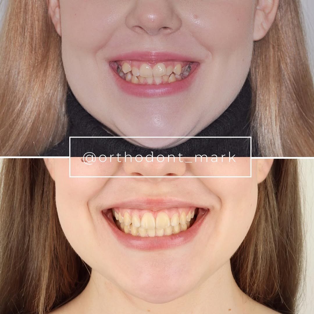
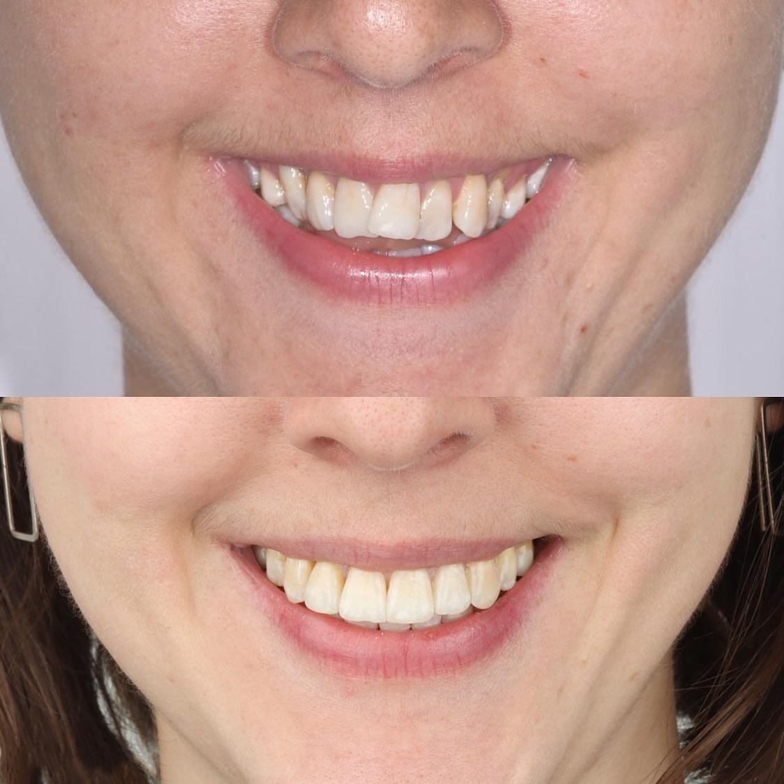
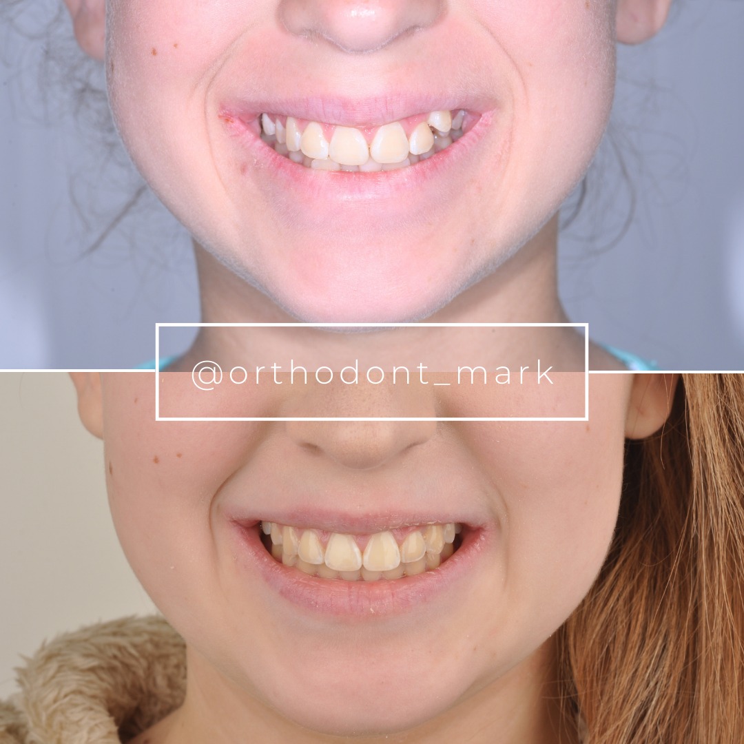
Clinical cases from Dr. Mark Radzhabov's practice
Learn the full MARPE protocol from Dr. Mark Radzhabov
Fundamental course covering CBCT patient selection, miniscrew planning, activation protocols, and 60+ clinical cases. Choose the access level that fits your practice.
Essentials of rapid palatal expansion for practicing orthodontists.
- Core RPE concepts and biomechanics
- 6 structured video lessons
- Clinical decision checklists
- Lifetime access to recordings
Deep-dive into MARPE protocol, diagnostics, and clinical execution.
- 6 modules covering MARPE from A to Z
- CBCT case selection walkthroughs
- Miniscrew placement and activation
- 60+ annotated clinical cases
- Direct Q&A with Dr. Mark Radzhabov
5-element medical consultation framework for dentists and orthodontists.
- Trust-building consultation protocol
- 5 lesson modules
- Templates for treatment plan delivery
- Works with any clinical specialty
Clinical FAQ
How do I differentiate transverse deficiency from arch-length crowding?
Compare a skeletal-width measurement with tooth-size and space calculations. A documented lack of skeletal width supports expansion. A space deficit with acceptable skeletal width directs treatment toward alignment, reduction, restorative planning, or other space management.
Which diagnostic criteria confirm a skeletal transverse deficit before MARPE?
Use palatal molar landmarks, occlusal assessment, arch form, and imaging when indicated. Sant et al. (2016) included skeletally mature patients with transverse maxillary skeletal deficiency greater than 5 mm.
When is palatal expansion unnecessary for dental crowding?
Palatal expansion is unnecessary when the diagnostic problem is inadequate arch perimeter or tooth-size discrepancy without evidence of skeletal constriction. Measure crown widths and available space before choosing an expansion appliance.
How do I distinguish maxillary constriction from arch-length discrepancy?
Maxillary constriction concerns skeletal width and transverse arch relationships. Arch-length discrepancy concerns whether tooth material exceeds usable arch space. Both can coexist, so one finding should not be used to assume the other.
Which measurements guide MARPE versus non-expansion treatment planning?
Dr. Radzhabov uses palatal sixth-tooth landmarks for McNamara assessment, mesiodistal crown measurements, and selected space analyses. ANS-PNS was used for maxillary horizontal diameter by Manabe et al. (2022).
Does intermolar width analysis establish a skeletal transverse deficiency?
No. Intermolar width is a useful screening measurement, but it must be interpreted with palatal anatomy, occlusion, crown dimensions, and skeletal assessment. A dental-width observation alone cannot establish the source of the discrepancy.
How should posterior crossbite diagnosis influence expansion selection?
Posterior crossbite should prompt evaluation of transverse skeletal width and dental inclinations. It is a clinical sign, not a standalone appliance prescription. The treatment choice depends on whether the underlying discrepancy is skeletal or dentoalveolar.
What does arch perimeter discrepancy mean in MARPE case selection?
Arch perimeter discrepancy describes a mismatch between tooth material and available arch space. It can explain crowding without proving a need for MARPE, so calculate it before attributing irregular alignment to maxillary constriction.
How does MSE expansion indications differ from crowding management?
MSE expansion indications require evidence of a skeletal transverse objective. Crowding management begins with crown-size and available-space analysis. Chun et al. (2022) reported similar dentoalveolar changes for MARPE and RPE except for maxillary width.
When should CBCT be considered in transverse orthodontic diagnosis?
Consider CBCT when skeletal anatomy or treatment response must be clarified beyond models and clinical examination. Lim et al. (2017) assessed MARPE with CBCT at T0, T1, and T2.
A defensible expansion decision begins with identifying the source of the discrepancy rather than reacting to aligned or crowded crowns. Dr. Mark Radzhabov’s approach places model calculations and landmark-based width assessment before appliance selection. Use a documented case review or enroll in structured training when the skeletal and dental findings conflict. Key sources: Manabe · 2022 · PLOS ONE · doi:https://doi.org/10.1371/journal.pone.0265272. Chun · 2022 · BMC Oral Health · doi:https://doi.org/10.1186/s12903–022-02138-w.