
Anchorage Load Sharing in Hybrid Expanders:
Force Distribution
Between Teeth and Bone Explained
Understand how documented appliance designs partition expansion force—and why that ratio predicts suture separation before activation begins.
TL;DR Anchorage load sharing in hybrid expanders governs how expansion force is divided between miniscrew-to-bone and band-to-tooth pathways. Suture maturation stage, patient sex, and bone density all modulate the effective skeletal response. Clinicians who understand these force-distribution mechanics can select appliance design more precisely and anticipate suture separation outcomes before activation begins.
When a clinician bonds a hybrid expander and begins activation, the force generated travels simultaneously through two distinct anatomical routes: cortical bone via palatal miniscrews, and the periodontal ligament via molar or premolar bands. How that load is partitioned between tooth and bone anchorage determines whether the outcome is true skeletal widening or predominantly dental tipping. Dr. Mark Radzhabov at ortodontmark.com examines the documented biomechanical principles and published imaging data behind hybrid expander load sharing, giving clinicians a framework for appliance selection and activation planning grounded in peer-reviewed evidence.
Why Does the Tooth-to-Bone Force Ratio Change
Suture Maturation
Between Patients of the Same Age?
Anchorage load sharing in hybrid expanders is the documented distribution of activation force between osseous anchorage units (palatal miniscrews) and dental anchorage units (molar or premolar bands) within a single palatal expansion appliance. The ratio is not fixed by the appliance geometry alone. It is continuously modulated by the resistance offered at each interface—and that resistance changes as the midpalatal and adjacent sutures mature. Understanding which sutures are closing, and at what rate, is therefore a prerequisite for predicting how an identical appliance will behave in two patients of nominally the same age. Govaerts et al. (2023) provided CBCT-based staging data that makes this variability concrete: pterygomaxillary suture closure ranges from 83% to 100% in females between ages 13 and 17 years. A patient at the lower end of that range offers substantially less osseous resistance posteriorly, which shifts a greater proportion of the activation force toward the anterior skeletal segment—an asymmetry that affects how the palatal vault opens and where the dental side effects concentrate. Clinicians relying on chronological age alone cannot detect this variation. Only high-resolution cross-sectional imaging reveals it. The midpalatal suture adds a further dimension. Govaerts et al. (2023) documented that midpalatal suture closure occurs in 61% of females at 15 years of age, while in the same population the pterygomaxillary suture may already be substantially obliterated. When the midpalatal suture is still partially open but the pterygomaxillary suture is resistant, hybrid expansion force is disproportionately absorbed at the posterior boundary, and the net forward vector of maxillary displacement may diminish. Recognizing this mismatch helps the clinician decide whether a tooth-borne component should be retained, reduced, or eliminated entirely from the appliance design.
How Do Sex-Based Differences in Suture Response
Redirect
the Anchorage Strategy for Hybrid Expansion?
The assumption that a given hybrid expander design produces equivalent skeletal effects across sexes is not supported by the published retrospective cohort data. Jeon et al. (2022) reported an overall MARPE suture separation success rate of 79.53%, with a breakdown that has direct implications for appliance selection: the rate reached 94.17% in females but only 61.05% in males. When one patient group approaches near-certainty of separation while another falls substantially below the three-quarter threshold, the optimum anchorage configuration is not the same for both, even when the CBCT suture stage looks similar. The age-by-sex interaction deepens this divergence. Jeon et al. (2022) found that older male patients showed statistically significant suture nonseparation, whereas females did not demonstrate this age-dependent effect. In practical terms, for male patients in mid-to-late adolescence, the hybrid expander design should weight the bone-borne component more heavily—additional miniscrew pairs, paramedian rather than lateral insertion sites, or a bone-borne-only protocol—because the tooth-borne component will not compensate for suture resistance that the miniscrews are failing to overcome. The female patient profile warrants different threshold-setting. For miniscrew-assisted rapid palatal expansion case selection, these figures reframe how the clinician counsels the patient and structures the activation schedule. A male patient whose CBCT shows a partially fused midpalatal suture presents a combination of elevated suture resistance and a statistically lower sex-specific separation probability. In that context, a hybrid expander with unreinforced dental anchorage is unlikely to generate the skeletal force vectors needed, and surgical assistance should enter the conversation earlier rather than later.
What Does the Discrepancy Between Device Activation
and Actual Separation
Reveal About Hybrid Force Dissipation?
An animal study by Klytong (2010) using micro-implant-assisted sutural expansion in rabbits quantified a gap that every clinician activating a hybrid expander should internalize: against a device activation of 5.6 mm, measured sutural separation ranged from 3.72 mm to 4.45 mm across experimental groups. The remaining force was absorbed elsewhere—by bony deflection, periodontal ligament compression, and viscoelastic deformation of surrounding craniofacial structures. This dissipation effect does not disappear in human patients. It scales with the magnitude of suture and osseous resistance present at the time of activation. For hybrid expander design, the Klytong (2010) data illustrates why increasing device activation beyond a calculated threshold does not proportionally increase skeletal separation. Once the tooth-borne component reaches its elastic limit at the molar support, further activation loads the miniscrews asymmetrically—and if miniscrew cortical engagement is insufficient, the excess force dissipates into the alveolar ridge rather than opening the suture. Clinicians designing the anchorage load sharing ratio should therefore treat the dental component as a ceiling, not a floor: it sets the maximum load the tooth route will tolerate before force is redirected entirely to bone. This dissipation principle also informs how practitioners interpret slow or stalled expansion. When a patient shows minimal arch width change after multiple activations, the first diagnostic question is not whether the screw is turning but where the force is going. CBCT taken mid-treatment to assess miniscrew position relative to cortical bone, combined with periodontal probing for dental tipping at the anchor teeth, can distinguish between suture resistance, miniscrew deflection, and appliance deformation—each of which requires a different corrective response.
How Did Onplant-Supported Protraction Establish
Bone-Borne Anchorage
as a Clinically Reliable Force Origin?
Before palatal miniscrews became the standard osseous anchor for expansion, the onplant—a disk-shaped subperiosteal implant—demonstrated that force applied directly to palatal bone could produce measurable skeletal displacement without dental intermediaries. Hong et al. (2005) reported a case in which a 7.7 mm onplant placed on palatal bone in an 11-year-old patient provided stable skeletal anchorage for maxillary protraction, achieving 2.9 mm of forward maxillary displacement. The relevance to hybrid expander mechanics is not the protraction vector itself but the confirmation that a relatively small-diameter osseous interface, when properly osseointegrated, can sustain clinically significant skeletal force without tooth involvement. The onplant case clarifies the theoretical justification for weighting the bone-borne component in contemporary hybrid expanders. When the dental component is reduced or eliminated, the entire activation force must be transferred through the osseous interface—which demands adequate cortical bone volume at the insertion site, confirmed angulation, and sufficient healing time before full loading. Hybrid expander designs that retain some dental anchorage represent a deliberate compromise: they accept a degree of dental side effect in exchange for reduced demand on each individual miniscrew. For clinicians exploring structured MARPE training who are deciding between a purely bone-borne MSE design and a hybrid hyrax with molar bands, the Hong et al. (2005) precedent is instructive: skeletal anchorage works without teeth, but the force per osseous unit rises accordingly. A four-miniscrew hybrid that also bands the first molars distributes peak load across six anchorage points. Removing the bands concentrates that same peak across four miniscrews alone. Which configuration is appropriate depends on the cortical bone density visible on CBCT and the suture maturation stage established before bonding.
Which CBCT Suture Findings Should Shift the Anchorage
Load Ratio
Toward Bone Before You Begin Activation?
Integrating the available imaging data into a practical decision framework begins with the pterygomaxillary suture. Because Govaerts et al. (2023) documented that pterygomaxillary closure ranges from 83% to 100% in females aged 13 to 17, a patient at the high end of that range is presenting a posterior osseous barrier that will substantially redirect activation force anteriorly. In that patient, retaining a full dental component may produce excessive loading of the anterior anchorage teeth with limited posterior skeletal benefit. Reducing the dental component and reinforcing the bone-borne pathway addresses the posterior barrier more directly. The SARPE referral threshold data from Govaerts et al. (2023) provides a useful calibration point: clinicians recommended surgical assistance at a cut-off of 15.1 years for orthodontists and 14.8 years for maxillofacial surgeons in female populations. These ages are not absolute limits but reflect the suture maturation stage at which non-surgical hybrid expansion becomes progressively less reliable. A female patient whose CBCT demonstrates advanced pterygomaxillary closure before age 15.1 may already warrant a surgical assistance conversation, regardless of chronological age—reinforcing that imaging stage, not birthdate, drives the anchorage prescription. Combining the sex-specific separation data from Jeon et al. (2022) with the suture staging data from Govaerts et al. (2023) gives the clinician a two-axis decision matrix: suture maturation on one axis, patient sex on the other. A female patient with a partially open midpalatal suture and a pterygomaxillary suture in the lower closure range is a strong candidate for hybrid expansion with standard tooth-to-bone load sharing. A male patient with a midpalatal suture showing dense interdigitation and a pterygomaxillary suture approaching full closure requires either a reinforced bone-borne design or surgical referral—the tooth-borne component will not rescue a biomechanically inadequate osseous result.
Free Clinical Guide
Which Expansion Appliance at Which Age?
Get Dr. Mark Radzhabov's guide to skeletal-expansion appliances — indications, pros & cons, and how to choose by patient age. Free to your inbox.
Prefer video? Watch the free demo lesson on MARPE appliance types, features and trade-offs.
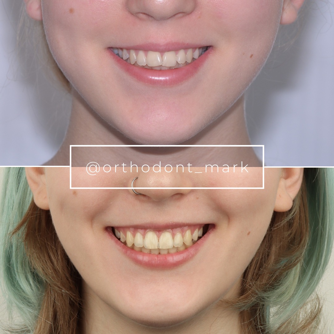
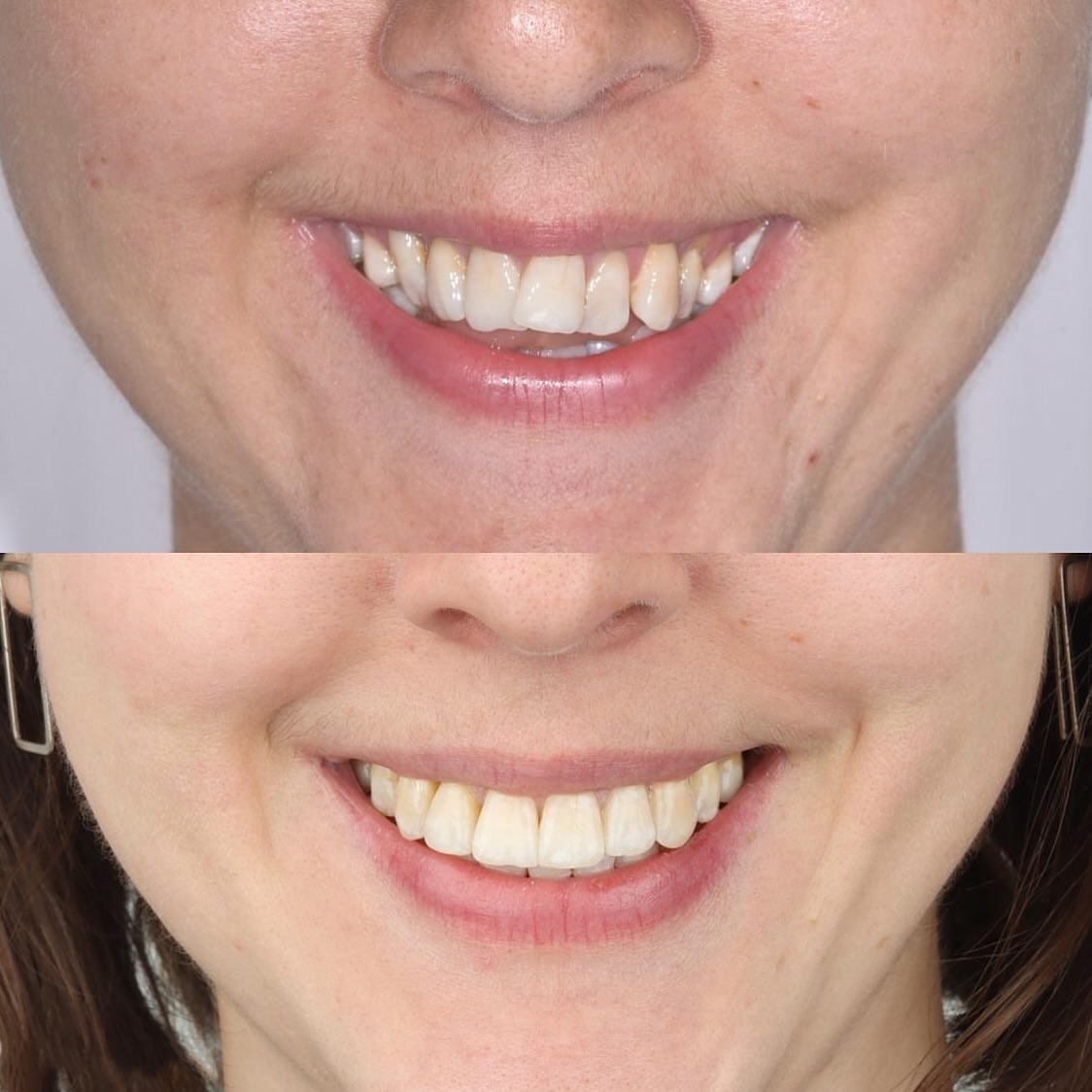
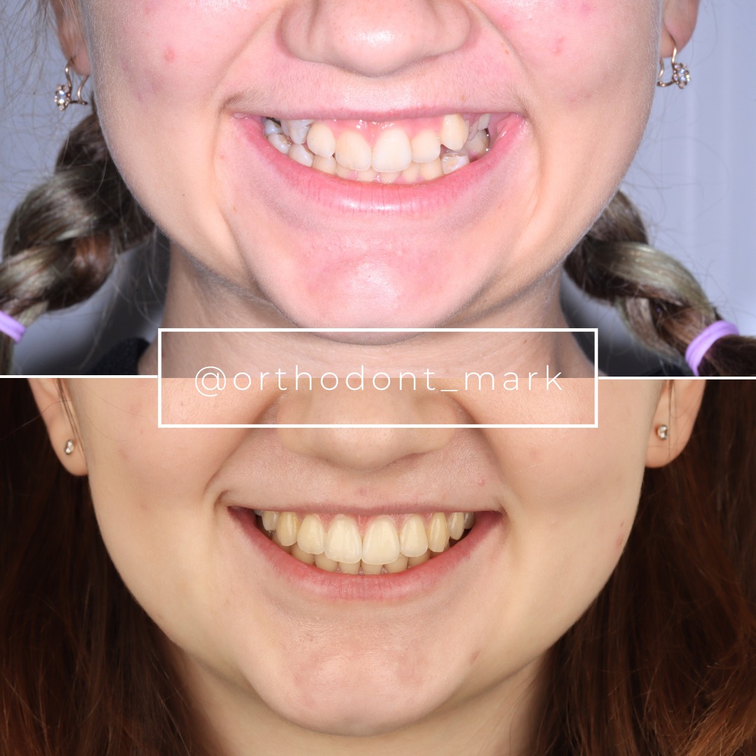
Documented outcomes
Clinical cases from Dr. Mark Radzhabov's practice
Learn the full MARPE protocol from Dr. Mark Radzhabov
Fundamental course covering CBCT patient selection, miniscrew planning, activation protocols, and 60+ clinical cases. Choose the access level that fits your practice.
Essentials of rapid palatal expansion for practicing orthodontists.
- Core RPE concepts and biomechanics
- 6 structured video lessons
- Clinical decision checklists
- Lifetime access to recordings
Deep-dive into MARPE protocol, diagnostics, and clinical execution.
- 6 modules covering MARPE from A to Z
- CBCT case selection walkthroughs
- Miniscrew placement and activation
- 60+ annotated clinical cases
- Direct Q&A with Dr. Mark Radzhabov
5-element medical consultation framework for dentists and orthodontists.
- Trust-building consultation protocol
- 5 lesson modules
- Templates for treatment plan delivery
- Works with any clinical specialty
Clinical FAQ
How does anchorage load sharing in hybrid expanders differ from a purely tooth-borne RPE?
A purely tooth-borne RPE routes all activation force through the periodontal ligament, producing predominantly dental tipping with limited skeletal effect. Hybrid expanders add palatal miniscrews so that a portion of force bypasses the teeth entirely and acts directly on the midpalatal suture, increasing true skeletal displacement.
Which CBCT suture finding most reliably predicts inadequate hybrid expansion force distribution in male patients?
Dense interdigitation at the midpalatal suture combined with high pterygomaxillary closure signals that osseous resistance will exceed what a standard hybrid design can overcome. Jeon et al. (2022) documented that older male patients show statistically significant suture nonseparation, making reinforced bone-borne anchorage or surgical referral the evidence-based response.
At what SARPE referral age should a clinician reconsider hybrid expander load sharing in female patients?
Govaerts et al. (2023) reported that orthodontists recommend surgical assistance at a cut-off of 15.1 years in females, and maxillofacial surgeons at 14.8 years. These thresholds reflect suture maturation stages where non-surgical hybrid load sharing becomes progressively unreliable, not absolute age-based surgical triggers.
How much of a hybrid expander's device activation is actually converted to sutural separation?
Klytong (2010) measured 3.72 to 4.45 mm of sutural separation against a device activation of 5.6 mm in a rabbit model. The remainder was dissipated by bony deflection and viscoelastic deformation. Clinicians should plan activation targets that account for this structural energy loss rather than assuming one-to-one conversion.
Does reducing the dental anchorage component in a hybrid expander improve skeletal outcomes?
Reducing the dental component concentrates force through the miniscrews, limiting dental tipping. However, Hong et al. (2005) demonstrated that purely osseous anchorage—even from a small 7.7 mm onplant—can achieve meaningful skeletal displacement of 2.9 mm. Adequate cortical bone depth and density must be confirmed by CBCT before eliminating dental anchorage entirely.
How does the pterygomaxillary suture closure range affect force distribution in adolescent female patients?
Govaerts et al. (2023) documented pterygomaxillary closure from 83% to 100% in females aged 13 to 17. Patients near the upper end of this range present a posterior osseous barrier that redirects hybrid activation force anteriorly, altering the arch widening vector and increasing anterior dental anchorage demand.
Is the midpalatal suture reliably open in female patients at age 15 for hybrid expansion planning?
Not reliably. Govaerts et al. (2023) found midpalatal suture closure in 61% of females at 15 years of age. Nearly two-thirds of 15-year-old females in that imaging study showed measurable closure, meaning CBCT staging is necessary—chronological age alone does not confirm suture patency for hybrid expansion.
How should hybrid expander anchorage design differ between male and female patients at the same skeletal age?
Jeon et al. (2022) showed overall MARPE suture separation of 79.53%, with females reaching 94.17% and males only 61.05%. For male patients, bone-borne anchorage weighting should be increased earlier—more miniscrew pairs or paramedian insertion—because the tooth-borne component alone does not compensate for the statistically lower separation probability.
What clinical signs indicate that hybrid expander force is routing through dental anchorage rather than the osseous pathway?
Progressive buccal crown tipping at banded molars, minimal measurable arch-width change on digital models, and diastema formation that stalls after early activation all suggest force dissipation through the periodontal ligament rather than suture opening. Mid-treatment CBCT confirming miniscrew position relative to cortical bone resolves the diagnosis.
When should a clinician refer to surgical assistance rather than optimizing hybrid expander load sharing further?
When CBCT shows advanced midpalatal and pterygomaxillary suture closure, the patient is male with age-related suture nonseparation risk per Jeon et al. (2022), and two activation cycles have produced no measurable sutural diastema, reconfiguring the hybrid design is unlikely to succeed and surgical assistance becomes the evidence-supported pathway.
Anchorage load sharing in hybrid expanders is not a static property of the device—it shifts with suture maturation stage, patient sex, and the proportion of force routed through miniscrew versus band. Clinicians who interpret CBCT suture staging before activation, and who adjust the tooth-to-bone force ratio accordingly, produce more predictable skeletal outcomes and limit unnecessary dental side effects. Dr. Mark Radzhabov encourages practitioners to bring specific cases for a structured review of imaging and appliance design at ortodontmark.com. Key sources: Govaerts et al. (2023), Journal of Orofacial Orthopedics, doi:10.1007/s00056–023-00487-x. Jeon et al. (2022), Clinical Oral Investigations, doi:10.1007/s00784–021-04281–0.