
MARPE Skeletal Anchorage Basics:
Force Redirection
Away From the Dentition
A peer-reviewed breakdown of how bone-borne expansion changes suture separation outcomes, dental side-effects, and case-selection thresholds for practicing orthodontists.
TL;DR MARPE skeletal anchorage basics center on routing expansion force directly through cortical bone, bypassing the dental roots entirely. Miniscrews anchored in the hard palate transmit load to the midpalatal suture rather than the crown-root complex. Chun et al. (2022) confirmed MARPE achieves a 95% suture separation rate versus 90% with conventional RPE, with less buccal tooth displacement.
When a patient presents with posterior crossbite and a crowded maxillary arch, the clinical question is not simply whether to expand — it is whether your force vector will move bone or tip teeth. Understanding MARPE skeletal anchorage basics is what separates a predictable skeletal result from a dentoalveolar compromise. Dr. Mark Radzhabov at ortodontmark.com has compiled the current controlled trial and cohort evidence to give practicing orthodontists a biomechanically grounded framework for choosing and loading palatal anchorage systems. This article covers the force pathway differences, suture separation outcomes, sex-related success variability, and the maturation signals that should redirect your protocol toward surgical assistance.
Why Does the Anchorage Source Change
What Moves?
MARPE skeletal anchorage is a palatal expansion approach in which titanium miniscrews placed into the cortical bone of the hard palate transmit activation forces directly to the midpalatal suture, eliminating the crown-root complex as an intermediary load-bearing element. In a conventional hyrax or bonded RPE, the entire activation load is transmitted through molar bands or acrylic pads resting on the maxillary premolars and molars. The teeth themselves become the structural link between the jackscrew and the suture, and that link exacts a biological cost: buccal crown tipping, alveolar bending, and periodontal strain accumulate before a single micron of sutural opening occurs. With palatal miniscrews — typically four units placed in a paramedian pattern straddling the mid-palatal raphe — the jackscrew frame bypasses dental roots entirely. The cortical plate of the hard palate becomes the primary load recipient, and the force vector arrives at the midpalatal suture with less mechanical attenuation through dentoalveolar structures. Chun et al. (2022) confirmed that MARPE produces less buccal displacement of anchor teeth during both the active expansion and consolidation phases compared to conventional RPE, a finding consistent with the biomechanical rationale above. The nasal floor and lateral nasal walls also respond differently under a bone-borne vector. Chun et al. (2022) documented greater nasal width increase at the molar region and at the greater palatine foramen with MARPE than with conventional RPE. This broader transverse remodeling reflects the more basal, less dental distribution of the applied force — a genuinely skeletal rather than dentoalveolar expansion pattern. For clinicians managing patients with nasal airway compromise alongside transverse deficiency, this differential nasal response carries direct treatment-planning significance. Explore the full miniscrew-assisted rapid palatal expansion protocol to understand appliance design choices that maximize this skeletal response.
How Reliably Does MARPE Split
the Suture,
and Who Is at Risk for Failure?
Suture separation — confirmed on CBCT as a triangular radiolucent gap widening from anterior to posterior — is the definitive radiographic proof that skeletal rather than dental expansion has occurred. Without that gap, any arch width gain is predominantly dentoalveolar and subject to greater relapse potential. Chun et al. (2022) reported a 95% midpalatal suture separation rate with MARPE compared to 90% with conventional RPE, a controlled-trial comparison that establishes a meaningful clinical advantage for the bone-borne design. Sex and skeletal maturation, however, modulate that advantage substantially. Jeon et al. (2022) found an overall MARPE suture separation rate of 79.53%, but the distribution across sexes diverged sharply: 94.17% in females versus 61.05% in males. This disparity is not simply an age artifact. Jeon et al. (2022) further demonstrated that older male patients showed significantly higher nonseparation rates with MARPE, whereas age did not significantly affect female outcomes in the same way — meaning suture fusion in males follows a distinct biological trajectory that chronological age alone cannot fully capture. For case selection, this evidence demands a sex-stratified approach. A male patient with CBCT signs of advanced suture interdigitation carries a materially higher risk of non-separation regardless of his stated age. Relying on a simple age threshold without imaging confirmation exposes the clinician to a predictable failure mode. Surgically assisted rapid palatal expansion becomes the more defensible choice once dense cortical bridging is visible across the posterior and middle thirds of the suture on axial CBCT slices — a threshold independent of sex or calendar year. Review conventional rapid palatal expander outcomes alongside MARPE data to contextualize these separation rates within your own case mix.
When Does Suture Maturation Override
Patient Age
as Your Decision Threshold?
Chronological age has long served as a proxy for skeletal readiness, but midpalatal suture morphology on CBCT provides a more direct and actionable signal. Govaerts et al. (2023) established that suture maturation becomes clinically significant as early as age 15 in females, with implications for which expansion modality is biomechanically appropriate. This finding challenges any protocol that reserves surgical referral exclusively for patients in their twenties or beyond. The practical implication: a 15-year-old female patient is not automatically a straightforward MARPE candidate simply because she falls within a conventionally accepted adolescent window. Govaerts et al. (2023) found that closed midpalatal sutures were present in 61% of females at age 15, a proportion large enough to make CBCT-based suture staging a clinical necessity rather than an optional upgrade. Without that imaging step, a substantial share of adolescent female patients would be expanded against a fused or near-fused suture, converting what should be a skeletal procedure into a predominantly dental one. For the practicing clinician, the decision algorithm should place CBCT suture staging before appliance selection — not as a retrospective check, but as the primary case-planning tool. Stage the suture using an axial slice at the level of the palatal vault, assessing the anterior, middle, and posterior thirds independently. Dense cortical bridging in the posterior third, even with a still-visible radiolucent line anteriorly, signals a high-risk profile for MARPE non-separation. Structured MARPE training and first-case mentorship can accelerate competence in reading these CBCT criteria before committing to a clinical protocol.
Does Bone-Borne Expansion Hold Its
Skeletal Gains
Through Consolidation?
Opening the midpalatal suture is one clinical milestone. Maintaining that sutural gap during the weeks of new bone deposition is an equally critical, and often underappreciated, phase. Conventional RPE designs transmit retention force through the same dental elements that transmitted the activation force, meaning any periodontal or root resorptive damage accumulated during expansion is then also loaded during retention. The MARPE design removes this compounding risk. Chun et al. (2022) demonstrated that MARPE achieves better maintenance of skeletal, dentoalveolar, and periodontal gains during the consolidation period compared to conventional RPE. This superior consolidation profile reflects the same biomechanical logic that drives the active-phase advantage: with force routed through osseous anchorage rather than dental crowns, the retentive load during consolidation does not place additional tipping or extrusive stress on the anchor teeth. The suture is held open by a device that rests against bone, and the surrounding periodontal ligament can recover undisturbed. For clinical protocol design, this finding argues for maintaining the MARPE appliance in full through the consolidation phase rather than transitioning early to a tooth-borne retainer. Premature removal of the bone-borne appliance before adequate mineralization of the sutural gap introduces a relapse vector at precisely the moment when new woven bone is least resistant to compressive load. The consolidation duration should be guided by CBCT evidence of new bone fill within the opened suture, not by a fixed calendar interval applied uniformly across all patients.
Free Clinical Guide
Which Expansion Appliance at Which Age?
Get Dr. Mark Radzhabov's guide to skeletal-expansion appliances — indications, pros & cons, and how to choose by patient age. Free to your inbox.
Prefer video? Watch the free demo lesson on MARPE appliance types, features and trade-offs.
Documented outcomes
Clinical cases from Dr. Mark Radzhabov's practice
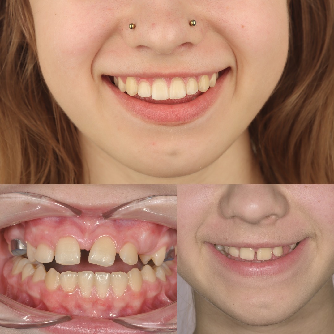
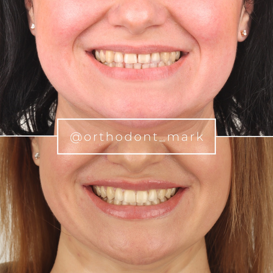
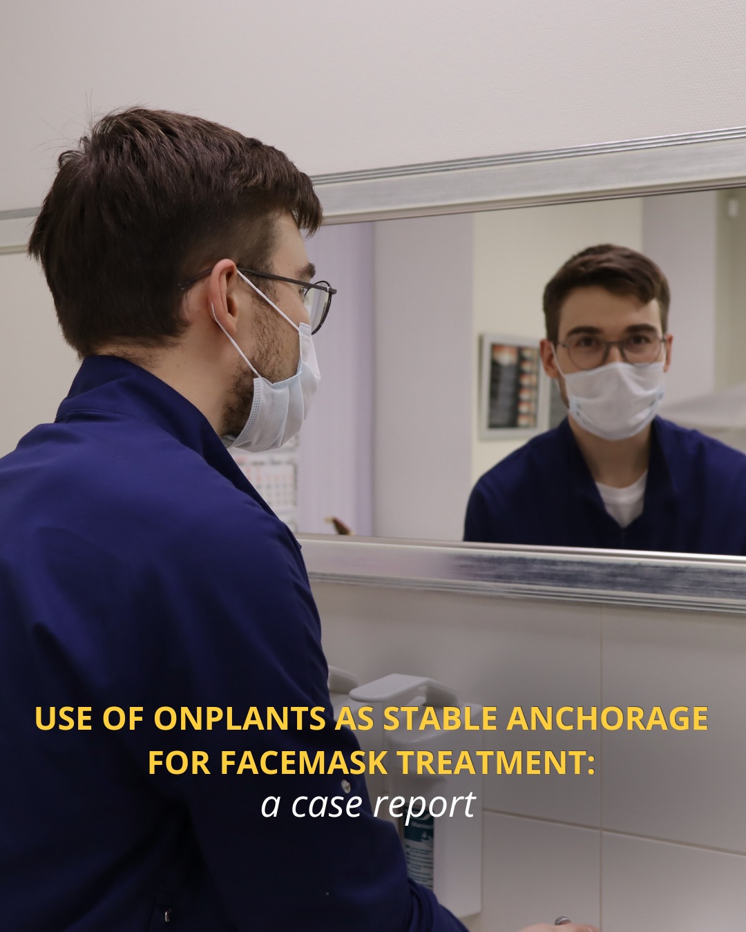
Learn the full MARPE protocol from Dr. Mark Radzhabov
Fundamental course covering CBCT patient selection, miniscrew planning, activation protocols, and 60+ clinical cases. Choose the access level that fits your practice.
Essentials of rapid palatal expansion for practicing orthodontists.
- Core RPE concepts and biomechanics
- 6 structured video lessons
- Clinical decision checklists
- Lifetime access to recordings
Deep-dive into MARPE protocol, diagnostics, and clinical execution.
- 6 modules covering MARPE from A to Z
- CBCT case selection walkthroughs
- Miniscrew placement and activation
- 60+ annotated clinical cases
- Direct Q&A with Dr. Mark Radzhabov
5-element medical consultation framework for dentists and orthodontists.
- Trust-building consultation protocol
- 5 lesson modules
- Templates for treatment plan delivery
- Works with any clinical specialty
Clinical FAQ
How does MARPE skeletal anchorage redirect expansion forces away from the dentition?
Paramedian palatal miniscrews anchor the jackscrew frame directly to cortical bone, so activation force travels from hard palate to midpalatal suture without passing through dental crowns or roots. This eliminates the crown-root complex as a load-bearing intermediate and reduces buccal tooth tipping.
What is the clinical advantage of bone-borne versus dental-borne palatal expansion force?
Chun et al. (2022) confirmed that directing force through osseous anchorage rather than molar bands produces less buccal anchor tooth displacement and better maintenance of skeletal gains during consolidation, alongside greater nasal width increase at the molar level and greater palatine foramen.
How does MARPE suture separation rate compare to conventional RPE in a controlled trial?
Chun et al. (2022) reported a 95% midpalatal suture separation rate with MARPE compared to 90% with conventional RPE, demonstrating a controlled-trial advantage for bone-borne anchorage in achieving true skeletal opening of the midpalatal suture.
Does sex affect MARPE suture separation success, and how should that influence case selection?
Yes. Jeon et al. (2022) documented a 94.17% separation rate in females versus 61.05% in males overall. Older male patients showed significantly higher nonseparation rates, making sex and CBCT suture morphology essential variables in pre-MARPE case assessment.
At what age does midpalatal suture maturation become clinically relevant for expansion planning?
Govaerts et al. (2023) found that suture maturation becomes clinically significant as early as age 15 in females, with closed sutures present in 61% of females at that age. This makes CBCT-based suture staging necessary in mid-adolescent patients, not just adults.
Should CBCT suture staging replace chronological age as the primary MARPE eligibility criterion?
Evidence supports staging suture maturity directly rather than relying on age. Govaerts et al. (2023) showed substantial suture fusion in teenage females, and Jeon et al. (2022) found age affected male but not female outcomes — confirming that imaging provides a more clinically actionable threshold than calendar age alone.
How long should the MARPE appliance remain in place during the consolidation phase?
The consolidation period should be guided by CBCT-confirmed new bone fill within the sutural gap rather than a fixed calendar interval. Chun et al. (2022) showed MARPE achieves better maintenance of skeletal and periodontal gains during consolidation than conventional RPE, supporting full appliance retention until imaging confirms adequate mineralization.
Which anatomical landmarks on CBCT best predict MARPE non-separation risk?
Dense cortical bridging in the posterior and middle thirds of the midpalatal suture on axial CBCT slices is the strongest imaging contraindication. The posterior third fuses earliest. A persisting anterior radiolucent line with posterior bridging does not indicate adequate suture patency for predictable non-surgical expansion.
Why does MARPE produce greater nasal width change than conventional RPE?
Because miniscrew anchorage applies force more basally through the palatal cortex, the remodeling envelope extends to the nasal floor and lateral nasal walls. Chun et al. (2022) measured greater nasal width increase in the molar region and at the greater palatine foramen with MARPE versus conventional RPE, reflecting a more apical skeletal expansion pattern.
When should surgical assistance be considered instead of MARPE for transverse maxillary deficiency?
Surgical referral is warranted when CBCT shows dense cortical bridging across the posterior and middle thirds of the midpalatal suture, particularly in male patients where Jeon et al. (2022) documented markedly lower MARPE success with advancing maturation. Sex-stratified suture imaging should drive that threshold, not age alone.
The biomechanical case for direct osseous anchorage in palatal expansion is now supported by controlled trial data showing superior suture separation rates and reduced dental side-effects compared with tooth-borne expanders. When you encounter a patient whose CBCT reveals advanced midpalatal suture fusion — particularly in males past adolescence — that imaging finding should recalibrate your protocol before a single miniscrew is placed. Dr. Mark Radzhabov offers structured case review and mentorship for orthodontists integrating MARPE into their workflow. Reach out through ortodontmark.com to discuss a current case or enroll in the next clinical training cohort. Key sources: Chun et al. (2022), BMC Oral Health, doi:10.1186/s12903–022-02138-w. Jeon et al. (2022), Clinical Oral Investigations, doi:10.1007/s00784–021-04281–0. Govaerts et al. (2023), Journal of Orofacial Orthopedics, doi:10.1007/s00056–023-00487-x.

