
MARPE Ankylosed Teeth Anchorage:
Force Redistribution
Rationale and Documented Outcomes
A peer-reviewed breakdown of bone-borne force vectors, suture separation predictors, and the imaging criteria that protect your protocol when posterior dentition is compromised.
TL;DR MARPE ankylosed teeth anchorage relies on palatal miniscrews that transmit orthopedic force directly to basal bone, bypassing compromised posterior dentition entirely. Bone-borne load distribution eliminates the buccal tipping and extrusion documented with tooth-borne systems. Sex and skeletal maturation remain the strongest predictors of suture separation success, as Jeon et al. (2022) and Govaerts et al. (2023) confirm.
When an ankylosed posterior molar sits within the proposed anchorage zone of a palatal expander, the standard tooth-borne protocol becomes biomechanically unreliable — force transmission is disrupted and dental side effects multiply unpredictably. MARPE ankylosed teeth anchorage addresses this directly: palatal miniscrews engage cortical bone independent of root support, redistributing orthopedic load across the midpalatal suture without relying on periodontally or structurally compromised teeth. Dr. Mark Radzhabov at ortodontmark.com examines the biomechanical rationale, the published success data, and the imaging criteria that allow clinicians to select and execute this protocol with confidence.
Why does tooth-borne expansion fail when an ankylosed molar
anchors
the appliance?
MARPE ankylosed teeth anchorage is a bone-borne palatal expansion strategy in which titanium miniscrews inserted into palatal cortical bone transmit orthopedic force directly to the midpalatal suture, decoupling load delivery from any ankylosed or structurally unreliable posterior teeth in the arch. An ankylosed molar has lost its periodontal ligament interface, meaning it cannot deform, tilt, or transmit elastic recoil the way a tooth with an intact PDL can. When a conventional hyrax or bonded RPE relies on that tooth as a primary abutment, force vectors become asymmetric, expansion stalls on the affected side, and reactive moments deflect into adjacent alveolar bone rather than opening the suture symmetrically. Bone-borne anchorage eliminates the undesirable molar movement and extrusion associated with tooth-borne expansion devices, a distinction documented by Hong et al. (2005) in palatal bone-borne appliance work. By anchoring entirely in the palate — at or near the anterior T-Zone described by Wilmes (2022) as the site of superior cortical bone quality — the clinician bypasses the ankylosed segment altogether. Force travels through the palatal vault, not through roots. The practical consequence is that buccal tipping of premolars and molars is significantly reduced compared to conventional RPE, as Chun et al. (2022) confirmed in a randomized controlled trial (p < 0.05). In a case complicated by ankylosis, that reduction matters even more: the clinician cannot depend on tooth-level compliance to self-correct asymmetric loading, so starting with a force path that avoids dental structures is not merely convenient — it is mechanically necessary.
Which patient factors most strongly predict suture separation
success
when posterior anchorage is compromised?
Selecting MARPE over a tooth-borne alternative only relocates the anchorage problem — it does not eliminate the biological challenge of opening a mature suture. Jeon et al. (2022) analyzed 215 subjects (95 males, 120 females) ranging from 6 to 60 years of age and found that overall MARPE suture separation success was 79.53%, but the distribution was strongly sex-dependent: females achieved 94.17% success versus 61.05% in males. Age compounds the male disadvantage. In males, increasing age is significantly associated with MARPE failure (p < 0.001), whereas in females this relationship does not reach significance (p = 0.221), per Jeon et al. (2022). For the clinician managing a male patient with an ankylosed molar, this means two independent risk factors converge: skeletal resistance at the suture and reduced force transmission through the remaining dentition. Both must be addressed in the consent discussion and the activation protocol. Skeletal maturation staging adds a further layer of case-selection discipline. Govaerts et al. (2023) found that at age 15 years, 61% of females already demonstrate closed midpalatal suture stages (D and E), placing the recommended orthodontic-to-surgical referral threshold at 15.1 years for orthodontists and 14.8 years for surgeons in females. In a patient with ankylosis who is already near this threshold, the combined risk profile — compromised dental anchorage, advancing suture fusion — should prompt early CBCT staging rather than a trial-and-error activation approach.
Where should miniscrews be placed when posterior
anchorage
teeth are ankylosed or absent?
Wilmes (2022) identifies the anterior palate T-Zone as the optimal miniscrew insertion site in MARPE based on superior cortical bone quality at that location. This recommendation becomes operationally critical when posterior teeth are ankylosed: the clinician cannot distribute anchorage distally toward compromised abutments without reintroducing tooth-borne force vectors, so the entire mechanical system must be supported anteriorly. The T-Zone — running from the incisive foramen region laterally toward the canine eminences — offers the thickest cortical layer accessible through the oral palate, providing primary stability for the miniscrews under sustained orthopedic load. Placing screws too far posteriorly, toward thinner palatal mucosa overlying the transverse palatine suture, reduces primary stability and risks screw mobility during the activation phase. In an ankylosis case, screw mobility translates directly into asymmetric or attenuated expansion force. Appliance design must then connect those anteriorly placed miniscrews to a rigid expansion frame that spans the arch without relying on the ankylosed tooth for structural support. Chun et al. (2022) demonstrated that MARPE achieves suture separation in 95% of cases compared to 90% with conventional RPE when identical expansion amounts are delivered — a margin that reflects the more direct skeletal loading pathway. When posterior dental anchorage is further compromised by ankylosis, the bone-borne architecture's advantage over tooth-borne designs only widens. For detailed placement protocols and case examples, review the MARPE skeletal expansion protocol and miniscrew biomechanics resource at ortodontmark.com.
How does pre-treatment CBCT imaging change the force-redistribution
decision
in ankylosis cases?
Confirming ankylosis itself requires CBCT: the absence of a periodontal ligament space around the affected root, combined with direct bone-to-root contact, distinguishes true ankylosis from severe hypercementosis or external resorption that could still permit limited tooth movement. Once the diagnosis is confirmed, the same imaging acquisition serves a second purpose — midpalatal suture maturation staging that determines whether MARPE or surgical expansion is the appropriate force strategy. Govaerts et al. (2023) found that 61% of females at age 15 already show stage D or E suture closure, with the recommended cut-off for switching from orthodontic to surgical protocols placed at 15.1 years for orthodontists. In a patient who presents with an ankylosed molar and is approaching this threshold, waiting for clinical signs of expansion failure before ordering CBCT is indefensible — staging must precede appliance fabrication, not follow it. Hounsfield unit mapping of palatal bone density at the proposed miniscrew sites further refines placement planning when posterior dental anchorage is absent. Denser cortical bone in the anterior palate tolerates higher initial insertion torque and resists the micro-movements that degrade primary stability during the activation phase. Clinicians considering submitting an ankylosis expansion case for consultation should include axial, coronal, and sagittal CBCT reconstructions through the full palatal vault alongside a periapical series of the ankylosed segment.
How does documented force redistribution in palatal expansion
protect
the ankylosed segment and adjacent teeth?
The central insight of bone-borne expansion in ankylosis management is that an ankylosed tooth, once removed from the force chain, stops being a problem and becomes a passive structural landmark. Hong et al. (2005) demonstrated that bone-borne anchorage eliminates the undesirable molar movement and extrusion seen with tooth-borne devices — a finding that extends logically to cases where the molar in question cannot move even if asked to, because ankylosis has eliminated its PDL-mediated compliance. Chun et al. (2022) confirmed in a randomized controlled trial that MARPE produces significantly less buccal tipping and displacement of anchor premolars and molars compared to conventional RPE (p < 0.05). In a patient with one or two ankylosed posterior teeth, this reduction in dental side effects across the remaining mobile dentition preserves the post-expansion occlusal platform and reduces the likelihood of relapse driven by tipped anchor teeth springing back toward their original positions. For the ankylosed tooth itself, the bone-borne protocol removes any risk of further root resorption, cemental disruption, or alveolar fracture that might accompany forceful tooth-borne loading against an immobile root. The expansion frame simply spans the vault above it, the miniscrews carry the load in cortical bone, and the suture receives symmetric force regardless of what lies between the two halves of the maxilla. Clinicians building this protocol into their practice workflow can access case-based teaching through structured MARPE training at ortodontmark.com.
Free Clinical Guide
Which Expansion Appliance at Which Age?
Get Dr. Mark Radzhabov's guide to skeletal-expansion appliances — indications, pros & cons, and how to choose by patient age. Free to your inbox.
Prefer video? Watch the free demo lesson on MARPE appliance types, features and trade-offs.
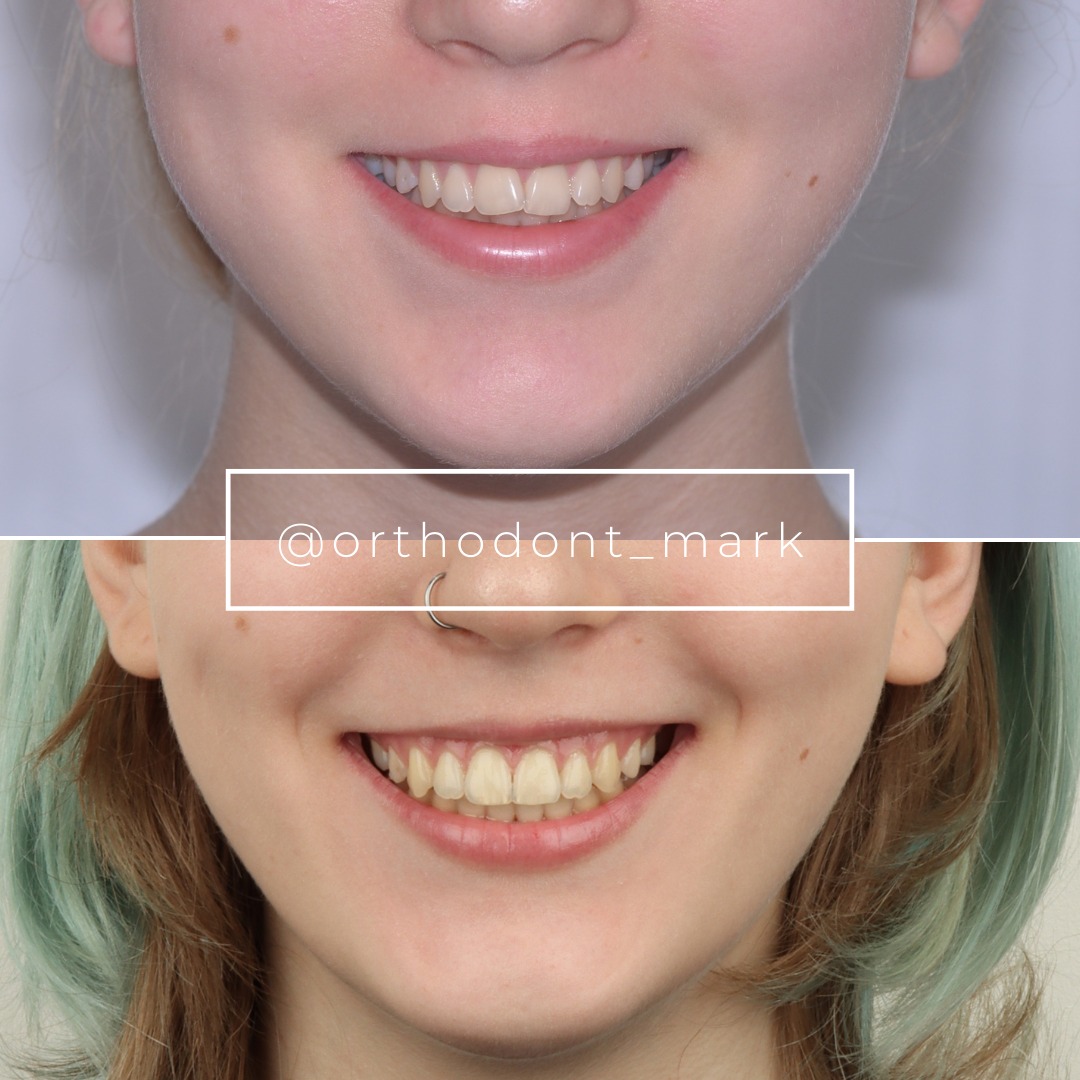
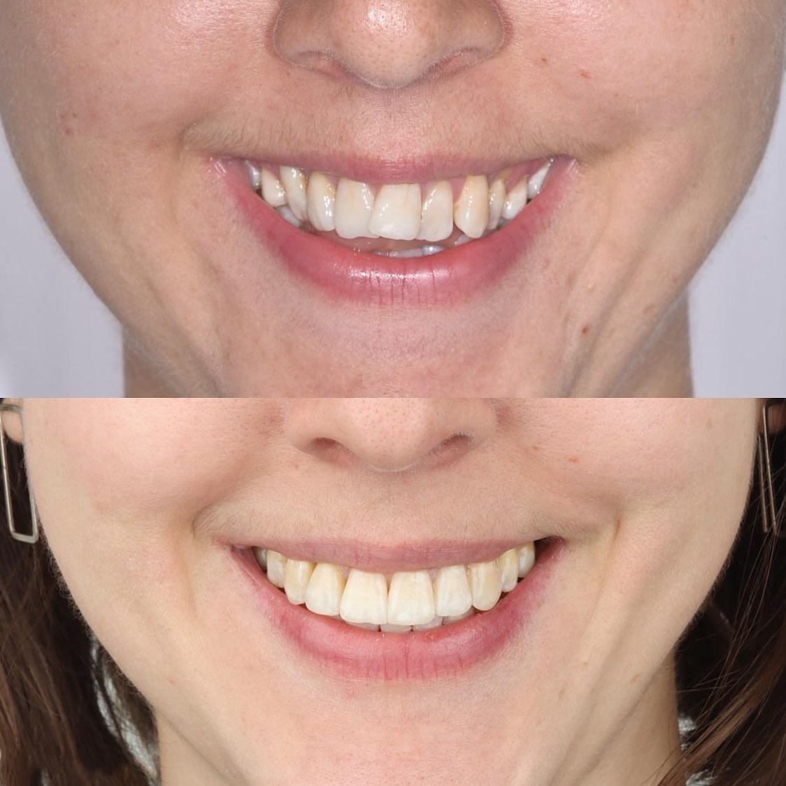
Documented outcomes
Clinical cases from Dr. Mark Radzhabov's practice
Learn the full MARPE protocol from Dr. Mark Radzhabov
Fundamental course covering CBCT patient selection, miniscrew planning, activation protocols, and 60+ clinical cases. Choose the access level that fits your practice.
Essentials of rapid palatal expansion for practicing orthodontists.
- Core RPE concepts and biomechanics
- 6 structured video lessons
- Clinical decision checklists
- Lifetime access to recordings
Deep-dive into MARPE protocol, diagnostics, and clinical execution.
- 6 modules covering MARPE from A to Z
- CBCT case selection walkthroughs
- Miniscrew placement and activation
- 60+ annotated clinical cases
- Direct Q&A with Dr. Mark Radzhabov
5-element medical consultation framework for dentists and orthodontists.
- Trust-building consultation protocol
- 5 lesson modules
- Templates for treatment plan delivery
- Works with any clinical specialty
Clinical FAQ
Can MARPE be used when an ankylosed molar occupies a planned anchor position?
Yes. Palatal miniscrews transfer force through cortical bone, bypassing the ankylosed tooth entirely. The ankylosed molar becomes a passive bystander rather than a failed abutment. Pre-treatment CBCT confirms ankylosis and guides miniscrew placement in the anterior T-Zone.
How does bone-borne anchorage change force redistribution in palatal expansion with ankylosis?
Bone-borne anchorage routes orthopedic load directly from miniscrews to the midpalatal suture, eliminating tooth-level intermediaries. Hong et al. (2005) documented that this eliminates undesirable molar movement and extrusion seen with tooth-borne devices, which is critical when a molar cannot transmit force due to ankylosis.
What CBCT criteria confirm true ankylosis versus severe hypercementosis before choosing a MARPE protocol?
True ankylosis appears on CBCT as complete absence of a PDL space with direct bone-to-root contact in axial and coronal reconstructions at high resolution. Hypercementosis shows an intact, if distorted, PDL space. Confirming ankylosis radiographically before appliance selection prevents a misdesigned force system.
Does patient sex affect MARPE suture separation success in cases complicated by posterior ankylosis?
Sex is a major predictor. Jeon et al. (2022) reported suture separation success of 94.17% in females versus 61.05% in males across 215 subjects. Male patients with ankylosed posterior teeth carry compounded risk: reduced dental anchorage and a lower baseline suture separation probability, particularly with advancing age.
At what suture maturation stage should a clinician abandon MARPE and refer for surgical expansion in an ankylosis case?
Stage D or E on midpalatal suture CBCT staging typically warrants a surgical referral. Govaerts et al. (2023) place the orthodontic-to-surgical threshold at age 15.1 years for orthodontists in females, with 61% of females already at stages D or E by age 15. Ankylosis adds further reason to avoid prolonged non-surgical trial.
Where should palatal miniscrews be inserted when posterior dental anchorage is unavailable?
Wilmes (2022) identifies the anterior palate T-Zone as the optimal site based on superior cortical bone quality. When posterior teeth are ankylosed or absent, all anchorage must be concentrated anteriorly. Posterior placement into thinner palatal bone risks screw mobility under sustained orthopedic load.
How much does MARPE reduce buccal tipping of anchor teeth compared to conventional RPE?
Chun et al. (2022) found in a randomized controlled trial that MARPE produces significantly less buccal tipping and displacement of anchor premolars and molars than conventional RPE (p < 0.05). In ankylosis cases, minimizing tipping of the remaining mobile dentition is especially important for post-expansion stability.
Does advancing age reduce MARPE success in male patients with posterior ankylosis?
Yes. Jeon et al. (2022) showed that increasing age is significantly associated with MARPE failure in males (p < 0.001) but not in females (p = 0.221). An older male presenting with posterior ankylosis therefore carries two independent risk factors and should be counseled accordingly, with surgical options discussed proactively.
What appliance design principles apply when fabricating a MARPE frame for a patient with posterior ankylosis?
The expansion frame must span the ankylosed segment without engaging it structurally. Miniscrews anchor exclusively in anterior palatal cortical bone per Wilmes (2022). The rigid frame distributes symmetric force across the suture. No band or bonded extension should rely on the ankylosed tooth for support or load transfer.
How does the MARPE suture separation rate compare to conventional RPE when used in complex ankylosis cases?
Chun et al. (2022) reported suture separation in 95% of MARPE cases versus 90% with conventional RPE under identical expansion conditions. Bone-borne force delivery likely accounts for the difference. In ankylosis cases that advantage is amplified because tooth-borne alternatives cannot reliably engage the compromised abutment.
Ankylosed posterior teeth need not eliminate palatal expansion from the treatment menu, provided the clinician shifts force delivery to a bone-borne architecture from the outset. Miniscrew placement in the anterior palate T-Zone, pre-treatment CBCT staging of the midpalatal suture, and careful attention to sex-dependent maturation patterns collectively define a defensible protocol. Dr. Mark Radzhabov has structured these principles into a clinically reproducible framework at ortodontmark.com — submit a case for review or enroll in structured MARPE training to apply this evidence at your next expansion appointment. Key sources: Jeon et al. (2022), Clinical Oral Investigations, doi:10.1007/s00784–021-04281–0. Govaerts et al. (2023), Journal of Orofacial Orthopedics, doi:10.1007/s00056–023-00487-x. Chun et al. (2022), BMC Oral Health, doi:10.1186/s12903–022-02138-w.