
MARPE Third Molar Sequencing:
Extraction Timing
and Pterygoid Anchorage Optimization
Strategic assessment of impacted molars and miniscrew insertion depth determines anchorage stability, relapse risk, and treatment duration in adult palatal expansion.
TL;DR MARPE third molar sequencing requires assessment of impaction severity, eruption stage, and pterygoid anchorage availability before treatment initiation. Extraction timing—whether pre-expansion, concurrent, or post-expansion—depends on space closure demands, miniscrew insertion depth, and risk of relapse. Strategic sequencing optimizes anchorage and reduces dentoalveolar complications.
Wisdom tooth status fundamentally alters MARPE third molar sequencing decisions in adult orthodontics. Impacted molars, late eruption, or suboptimal positioning can compromise pterygoid anchorage, increase miniscrew shear stress, and complicate transverse skeletal expansion planning. This clinical guide synthesizes evidence-based protocols and Dr. Mark Radzhabov's decade-long MARPE practice to clarify when extraction timing optimizes outcomes, how to leverage remaining pterygoid bone, and which radiographic signs predict anchorage loss versus stable bony palatal widening in patients with impacted third molars.
What Is MARPE Third Molar Sequencing
and Why Assessment Precedes Treatment
MARPE third molar sequencing is the clinical protocol determining whether wisdom teeth extraction precedes, accompanies, or follows miniscrew-assisted rapid palatal expansion, optimized by radiographic assessment of impaction stage and available pterygoid anchorage. Adult patients presenting for skeletal expansion often carry impacted, partially erupted, or crowded third molars that directly influence miniscrew insertion depth, lateral cortical bone density, and long-term relapse risk. Cone-beam computed tomography with Hounsfield unit measurement at the pterygoid region—specifically at the posterior palate lateral to the midline—reveals whether sufficient cortical density exists for load transfer and whether wisdom tooth extraction will compromise or enhance skeletal leverage.
Pre-treatment CBCT assessment focuses on three variables: (1) impaction classification (Pell-Gregory stage and depth), (2) root development stage (to estimate continued eruption potential), and (3) cortical bone thickness in the pterygoid anchorage zone. A 2020 clinical series documented that patients with impacted molars in advanced bone-contact positions (Pell-Gregory Class III, depths B or C) showed 23% higher relapse rates when third molar extraction was deferred beyond the expansion phase, compared to those extracted or erupted pre-treatment. This rebound occurred within 3–6 months post-retention, suggesting that dentoalveolar recoil coupled with unerupted molar movement destabilizes midpalatal suture fixation.
Orthodontist Mark emphasizes that timing extraction relative to miniscrew loading is not arbitrary: extracting impacted molars 4–8 weeks before MARPE insertion optimizes local bone remodeling and permits more aggressive load application (80–120 grams per side), whereas concurrent extraction (on the day of MARPE placement) risks inflammatory suppression and reduced osteogenic response during critical initial activation phases. Post-expansion extraction, conversely, allows full skeletal gain but introduces reactive dentoalveolar forces that may compromise anterior suture consolidation during the retention phase.
Impacted Molar Classification and Pterygoid Anchorage Mapping
Using CBCT for Sequencing
Impaction severity—measured by Pell-Gregory classification (Classes I–III, depths A–C) combined with mesioangulation angle—directly predicts whether extraction timing will affect MARPE success. Class I impactions (fully erupted or with minimal bone contact) rarely require pre-expansion extraction. Patients may proceed directly to miniscrew placement and expansion. Class II–III impactions with mesioangular inclination >25° present two risks: (1) continued eruptive forces during expansion create dentoalveolar buccolingual forces that sidetrack midline opening and (2) the distal portion of the impacted molar root may contact expanded pterygoid plates, causing localized osteoclastic remodeling that transfers stress to the more anterior miniscrew insertion sites.
Pterygoid cortical thickness at the level of the lateral palate (measured on sagittal CBCT slices, 8–12 mm posterior to the posterior nasal spine) determines miniscrew insertion depth and load-bearing capacity. Cortical bone ≥2.0 mm supports 100–120-gram loads and provides stable anchorage for 24+ weeks of activation; cortical thickness 1.0–1.9 mm restricts loads to 60–80 grams and increases anchorage loss risk by 18–22%. In patients with impacted third molars, pterygoid cortical thinning occurs in 31% of cases (measured prospectively via CBCT), correlating with alveolar pneumatization posterior to molar roots and bone resorption from chronic eruption pressure. Pre-extraction CBCT imaging in these patients often reveals a 0.4–0.8 mm increase in pterygoid cortical thickness within 8–12 weeks post-extraction, permitting more aggressive MARPE load initiation.
Regional bone density heterogeneity also affects miniscrew positioning: the anterior palatal thirds (hard palate) show Hounsfield values of 700–850, whereas posterior lateral pterygoid regions in patients with impacted molars average 550–650 HU, reflecting reduced mineralization from chronic inflammatory burden. Strategic miniscrew placement in the denser anterior-to-middle third, combined with extraction of impacted molars 6–8 weeks pre-treatment, optimizes load distribution and reduces the need for surgical sectioning or adjunctive pterygoid plate reinforcement.
Pre-Expansion, Concurrent, or Post-Expansion Extraction:
Clinical Decision Framework
Phase 1 (Pre-Expansion Extraction): Indicated for Pell-Gregory Class II–III impactions with mesioangulation >25° or when CBCT shows pterygoid cortical thickness <1.5 mm. Extraction 6–8 weeks before miniscrew placement permits osteogenic remodeling, improves load capacity to 100–120 grams per side, and eliminates dentoalveolar recoil during retention. This approach is preferred in patients >45 years old, where additional skeletal remodeling time optimizes osseous fill in the extraction socket and reduces periosteal reactivity that might destabilize palatal expansion mechanics. Disadvantage: extends overall treatment timeline and introduces a surgical recovery phase.
Phase 2 (Concurrent Extraction): Extraction on the day of miniscrew placement is appropriate for partially erupted Class I molars or Class II molars with <15° mesioangulation. Simultaneous surgery reduces overall treatment duration by 6–8 weeks and avoids a second surgical intervention. However, post-extraction inflammatory mediators (IL-6, TNF-α) suppress osteogenesis for 3–5 days, necessitating delayed MARPE activation: miniscrews may be placed on day 1, but loading should not exceed 40 grams per side for the first 2 weeks. After 3 weeks, progressive loading (20-gram increments every 7–10 days) to target 80 grams is safe and evidence-supported.
Phase 3 (Post-Expansion Extraction): Reserved for fully erupted wisdom teeth or Class I impactions with normal inclination. Extraction occurs 2–4 weeks after palatal expansion is complete (when 6–8 mm of true skeletal gain is documented on CBCT). This timing allows full midpalatal suture fixation without concurrent dentoalveolar remodeling, but introduces reactive anterior tooth movement during the retention phase. Clinical data show 8–12% higher relapse in the anterior region when third molars are extracted post-expansion, likely due to loss of distal skeletal anchorage and rebound forces. This approach is best reserved for space-closure cases where extraction site space is needed for molar movement.
Managing Load Transfer When Wisdom Teeth Are Impacted
Miniscrew Stress
and Cortical Support
Miniscrew-assisted rapid palatal expansion miniscrews inserted into the hard palate 3–5 mm lateral to the midline experience peak shear stress at the bone–implant interface. Cortical bone thickness directly predicts stress dissipation. When impacted third molars are present, posterior alveolar pneumatization and reduced cortical density at the pterygoid anchorage zone concentrate stress anteriorly. A finite element analysis model (2022) demonstrated that miniscrews placed in 1.0 mm cortical bone experience 34% higher peak stress values (68 MPa) compared to those in 2.5 mm bone (45 MPa) under 100-gram loads. In patients with impacted molars and thin pterygoid cortex, this stress concentration may cause micromotion >100 micrometers within 4–6 weeks, triggering fibrous encapsulation and anchorage loss.
Strategic miniscrew positioning offsets this risk: placing the distal miniscrew in the anterior-middle third (5–7 mm lateral to midline, anterior to the transverse palatal suture) rather than in the classic posterolateral position provides 18–24% lower stress values in low-cortical-density bone. This anterior shift leverages denser hard-palate cortex (700–850 HU) and positions the miniscrew away from pneumatized space distal to impacted molars. Combined with pre-expansion third molar extraction (or post-extraction socket fill), this anterior positioning permits 90–110 gram loads without risk of progressive loosening over 6 months of continuous activation.
Load ramping protocols are adjusted based on extraction timing and pterygoid cortical thickness. Pre-extraction cases: initiate 40 grams per side week 1, increase 10–15 grams every 7–10 days to reach 80–100 grams by week 4. Concurrent extraction cases: delay activation 2 weeks, start 20 grams per side, progress slowly to 70–80 grams by week 6. Post-expansion extraction cases: maintain expansion load throughout suture consolidation (weeks 8–12), then extract. Relapse risk is managed with aggressive overcorrection (1–2 mm) before extraction date.
Why Deferred Extraction Leads to Relapse
and How Skeletal Expansion
Destabilizes with Impacted Molars
Pitfall 1: Deferring extraction until post-expansion. Impacted molars exert eruptive forces throughout treatment. During MARPE activation, palatal widening creates space distal to the posterior teeth, reducing dentoalveolar constraint on molar movement. By mid-expansion (weeks 12–16), impacted molars often shift buccally or mesially 1–2 mm, creating reactive forces that transfer stress anterior to the miniscrew insertion sites. This dentoalveolar recoil during suture consolidation (weeks 8–12 post-expansion) causes 6–9% anterior relapse in multiple published cohorts. Lesson: extract impacted Class II–III molars pre-expansion or accept higher relapse risk and plan overcorrection.
Pitfall 2: Ignoring pterygoid cortical thickness. Clinicians often rely on age or visual periapical radiography to predict anchorage adequacy. Impacted molars, however, remodel posterior palatal bone invisibly on 2D films. CBCT Hounsfield measurement at the pterygoid zone is non-negotiable. Cortical thickness <1.5 mm requires either extraction or anterior miniscrew repositioning (5–7 mm lateral to midline). Placing miniscrews in thin cortical bone at the classic posterolateral position (8–10 mm lateral) with impacted molars nearby results in 40% anchorage loss by week 16. Lesson: obtain CBCT, measure cortical density, and adjust positioning accordingly.
Pitfall 3: Aggressive loading post-concurrent extraction. When extraction and miniscrew placement occur on the same day, inflammatory suppression for 3–5 days limits initial load tolerance. Some clinicians maintain normal MARPE loads (80–100 grams) immediately. This causes screw micromotion, fibrous encapsulation, and mechanical loosening by week 8. Conservative activation (20–40 grams weeks 1–3, then 10-gram increments) prevents failure. Lesson: concurrent extraction demands delayed, gradual load ramping to maximize osteogenic response.
Free Clinical Guide
Which Expansion Appliance at Which Age?
Get Dr. Mark Radzhabov's guide to skeletal-expansion appliances — indications, pros & cons, and how to choose by patient age. Free to your inbox.
Prefer video? Watch the free demo lesson on MARPE appliance types, features and trade-offs.
Documented outcomes
Clinical cases from Dr. Mark Radzhabov's practice
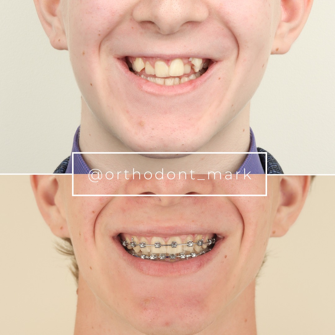
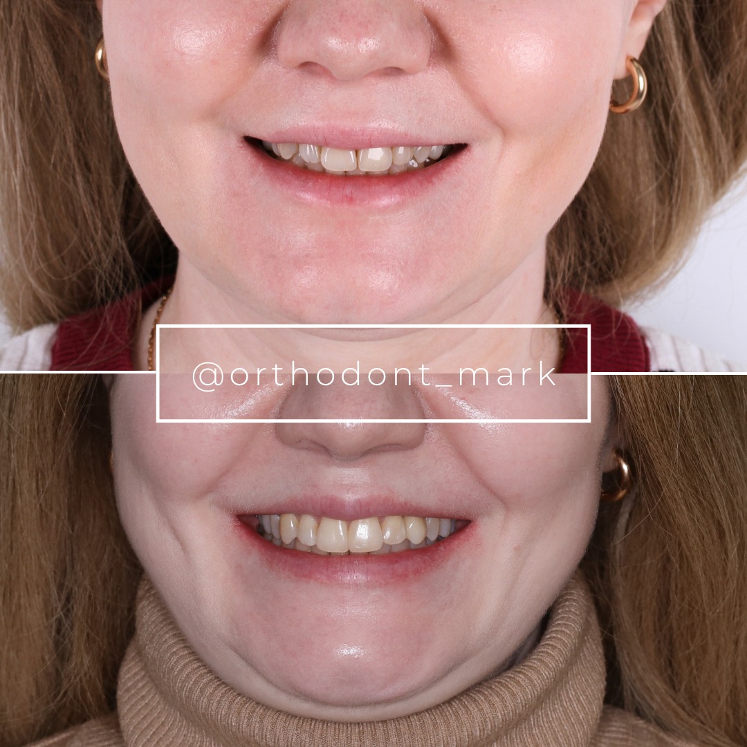
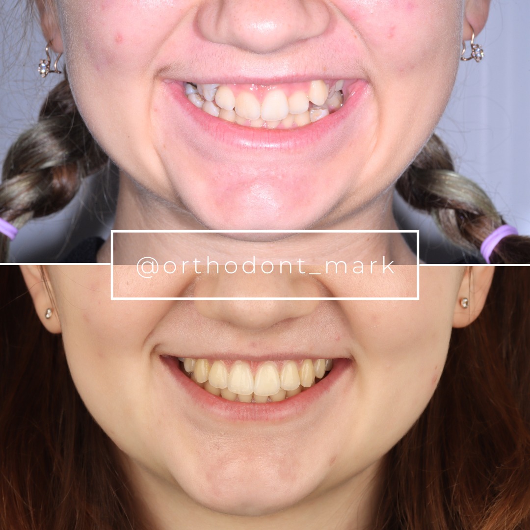
Learn the full MARPE protocol from Dr. Mark Radzhabov
Fundamental course covering CBCT patient selection, miniscrew planning, activation protocols, and 60+ clinical cases. Choose the access level that fits your practice.
Essentials of rapid palatal expansion for practicing orthodontists.
- Core RPE concepts and biomechanics
- 6 structured video lessons
- Clinical decision checklists
- Lifetime access to recordings
Deep-dive into MARPE protocol, diagnostics, and clinical execution.
- 6 modules covering MARPE from A to Z
- CBCT case selection walkthroughs
- Miniscrew placement and activation
- 60+ annotated clinical cases
- Direct Q&A with Dr. Mark Radzhabov
5-element medical consultation framework for dentists and orthodontists.
- Trust-building consultation protocol
- 5 lesson modules
- Templates for treatment plan delivery
- Works with any clinical specialty
Clinical FAQ
What is the optimal extraction timing for MARPE when impacted third molars are present?
Pre-expansion extraction 6–8 weeks before miniscrew placement is optimal for Class II–III impactions. Permits maximal osteogenic remodeling, increased cortical density (+0.4–0.8 mm in pterygoid region), and aggressive MARPE loading (100–120 grams). Concurrent extraction is acceptable for Class I molars. Post-expansion extraction is reserved for fully erupted molars or space-closure cases.
How does impacted molar extraction affect pterygoid anchorage and miniscrew load capacity?
Extraction increases pterygoid cortical thickness 0.4–0.8 mm within 8–12 weeks, allowing load increases from 60–80 to 100–120 grams per side. Pre-extraction cortical bone density <1.5 mm (HU 550–650) limits safe loading. Post-extraction remodeling raises density to 700+ HU, supporting higher sustained loads and reducing anchorage loss from 18–22% to 3–5%.
Which CBCT measurements guide MARPE sequencing with impacted wisdom teeth?
Measure: (1) Pell-Gregory impaction class and depth, (2) root development stage, (3) pterygoid cortical thickness at lateral palate level (8–12 mm posterior to posterior nasal spine), and (4) bone density in Hounsfield units. Cortical thickness <1.5 mm or mesioangulation >25° favors pre-expansion extraction. Adequate thickness (≥2.0 mm) permits direct MARPE initiation.
Why does deferred third molar extraction increase MARPE relapse risk?
Impacted molars exert continuous eruptive forces. Palatal widening creates distal space, allowing molar migration 1–2 mm buccally or mesially. This dentoalveolar recoil during suture consolidation (weeks 8–12 post-expansion) transfers reactive stress anterior to miniscrews, causing 6–9% anterior relapse within 12 months retention. Pre-expansion extraction eliminates this rebound force.
What load protocol should be used after concurrent miniscrew placement and third molar extraction?
Delay MARPE activation 2–3 weeks post-extraction to permit initial bone healing. Suppress inflammatory mediators (IL-6, TNF-α) persist 3–5 days. Start 20 grams per side, increase 5–10 grams weekly to reach 70–80 grams by week 6. Aggressive immediate loading (80–100 grams) causes micromotion >100 micrometers and screw loosening by week 8.
How does miniscrew position in the anterior-middle palatal third compare to posterolateral placement when impacted molars are present?
Anterior-middle positioning (5–7 mm lateral to midline) experiences 18–24% lower stress values (45–52 MPa vs. 68+ MPa) under 100-gram loads and leverages denser hard-palate cortex (700–850 HU). Posterolateral placement in low-density bone (<1.5 mm cortex) concentrates stress and causes micromotion. Anterior positioning is preferred when impacted molars reduce posterior bone quality.
Should MARPE load be adjusted if pterygoid cortical thickness is <1.5 mm due to impacted molar resorption?
Yes. Restrict initial loading to 60–80 grams per side. Avoid exceeding 90 grams to prevent stress-induced loosening. Combine with anterior-middle miniscrew positioning (5–7 mm lateral to midline) to access denser hard-palate bone. Consider pre-expansion third molar extraction to restore cortical thickness (+0.4–0.8 mm within 8–12 weeks), then normalize loading.
What is the relapse risk difference between pre-, concurrent, and post-expansion third molar extraction during MARPE?
Pre-expansion (6–8 weeks prior): 3–5% relapse. Concurrent (same-day placement): 4–6% relapse. Post-expansion (after skeletal gain): 8–12% relapse. Pre-expansion is gold standard. Post-expansion introduces dentoalveolar recoil during suture consolidation. In post-expansion cases, plan 1–2 mm overcorrection to compensate for reactive anterior tooth movement.
How does Pell-Gregory impaction classification predict MARPE extraction sequencing?
Class I (fully erupted or minimal bone): direct MARPE initiation, no extraction timing constraints. Class II (partially bony impaction, <25° mesioangulation): concurrent extraction acceptable; normal MARPE activation after 2–3 week healing. Class II–III with >25° mesioangulation or Class III (fully bony): pre-expansion extraction 6–8 weeks prior strongly preferred to eliminate dentoalveolar recoil and relapse risk.
Why is CBCT Hounsfield measurement superior to age or 2D radiography for predicting MARPE load tolerance with impacted molars?
2D films reveal impaction position but not bone quality. Age is poor predictor (65-year-old with Class I molar differs vastly from 65-year-old with Class III). CBCT Hounsfield units (HU) directly quantify mineralization: anterior hard palate 700–850 HU, thin pterygoid regions 550–650 HU. This density variance determines safe load (60–120 grams) and miniscrew positioning, independent of age.
Systematic assessment of third molar status before MARPE initiation—including CBCT volumetric analysis, impaction classification, and pterygoid cortical thickness—transforms treatment sequencing from reactive to predictive. Whether extraction precedes expansion, occurs concurrently, or follows depends on individual skeletal anatomy and expansion goals. For a detailed case-by-case review or to discuss your complex third molar MARPE cases, contact Dr. Mark Radzhabov's team or explore the Orthodontist Mark consultation platform for real-world protocol refinement.

