
MARPE Facemask Timing Class III
Sequencing Evidence for Late Adolescents
Evidence-based sequencing criteria for combining skeletal expansion and protraction in late-adolescent Class III constriction cases.
TL;DR Combined MARPE and facemask protraction timing class III constriction treatment requires deliberate sequencing in late adolescents. Chun et al. (2022) found suture separation in 95% of MARPE patients, confirming adequate maxillary mobility before protraction forces are applied. Asymmetric expansion and gingival complications must be monitored throughout, as they directly influence when protraction can safely begin.
Sequencing maxillary expansion with facemask protraction in late adolescents with Class III constriction remains one of the most consequential clinical decisions an orthodontist faces. Get the timing wrong and protraction forces meet a structurally inadequate maxilla, compromising both sagittal correction and skeletal stability. Dr. Mark Radzhabov at ortodontmark.com examines the published evidence on combined MARPE and facemask protraction timing, drawing on peer-reviewed trials and imaging studies to help clinicians build a defensible, reproducible protocol for this challenging patient group.
Why Does Suture Status Determine When Protraction
Begins
in Late Adolescents?
Combined MARPE and facemask protraction is a two-stage skeletal correction strategy in which bone-borne maxillary expansion achieves midpalatal suture separation before orthopedic protraction forces are applied to the mobilized maxilla. The sequencing logic rests on a biomechanical premise: a sutural split reduces circummaxillary resistance, theoretically making the maxilla more responsive to anterior traction. Applying protraction forces before confirmed separation risks loading a rigid structure and displacing teeth rather than the skeletal base. Chun et al. (2022) demonstrated suture separation in 95% of MARPE patients, compared with 90% of those treated with conventional RPE — a finding with direct implications for sequencing decisions. The higher separation rate with bone-borne mechanics suggests MARPE is a more reliable precursor to protraction in this age group, where suture patency is already narrowing. Clinicians relying on the miniscrew-assisted rapid palatal expansion protocol should treat this separation benchmark as a prerequisite gate, not an assumed outcome. Imaging confirmation of suture separation — typically a radiolucent diastema visible on periapical or CBCT imaging — marks the transition point between expansion and protraction phases. Without that confirmation, the clinician cannot distinguish dental tipping from true skeletal widening, and protraction forces added prematurely will act on a mechanically disadvantaged maxilla. The evidence supports a deliberate pause between phases rather than overlapping appliance activation.
Which Late Adolescents Are Poor Candidates for
Sequential
Expansion-Protraction?
Not every late adolescent with Class III constriction is an equally viable candidate for the combined protocol. Jeon et al. (2022) found that older age was significantly associated with MARPE suture separation failure in males but not in females — a sex-specific finding that reframes candidate screening. For older male adolescents approaching skeletal maturity, the probability of achieving the suture separation prerequisite is meaningfully lower, making the expansion phase the highest-risk link in the chain. This sex-based divergence has a practical implication: chronological age alone is insufficient for candidate selection. Skeletal maturation indicators — cervical vertebral staging or hand-wrist radiographs when indicated — must be layered onto the clinical picture, particularly for male patients in the upper adolescent age range. A male patient with advanced sutural ossification who fails expansion will never reach the protraction phase with adequate maxillary mobility. Female patients in the same cohort did not show the same age-related separation risk (Jeon et al., 2022), suggesting the suture remains more responsive in older female adolescents. Clinicians designing a class III constriction correction protocol should stratify male and female candidates separately rather than applying a single age cutoff to the entire group. This stratification should be documented in the consent and treatment-planning record.
How Should Clinicians Manage MARPE Complications
Before
Protraction Forces Are Added?
Introducing protraction forces while active MARPE complications are unresolved compounds patient burden and may compromise cooperation with the facemask — which demands consistent wear to be effective. Yoon et al. (2022) identified gingival inflammation around the MARPE appliance as the most prevalent complication, occurring in 83.9% of patients. That prevalence is high enough that peri-miniscrew soft-tissue management should be a standard part of the expansion phase rather than a reactive measure. Pain during or after expansion was reported by 45% of patients in the same analysis (Yoon et al., 2022), which has direct relevance to sequencing: a patient already managing expansion discomfort is less likely to achieve adequate facemask wear hours. Staggering the introduction of protraction — waiting until pain levels have normalized after the active expansion phase — is clinically rational even if it lengthens the overall timeline. Asymmetric expansion exceeding 1 mm occurred in 47.8% of MARPE patients (Yoon et al., 2022), a figure that should prompt midcourse CBCT evaluation before protraction begins. Asymmetric skeletal widening creates an unequal force platform for protraction, potentially producing a canted occlusal plane or asymmetric sagittal correction. Confirming bilateral suture separation and acceptable symmetry before activating facemask mechanics is a straightforward risk-reduction step supported by this complication rate.
What Is the Evidence-Based Checklist Before Initiating
Facemask
Protraction After MARPE?
A structured go/no-go checklist between phases reduces the risk of loading a maxilla that has not achieved adequate skeletal mobility. The four criteria below synthesize the findings reviewed and reflect the complication prevalence data from Yoon et al. (2022) and the separation benchmarks from Chun et al. (2022). First, confirm radiographic suture separation — a visible midpalatal lucency on periapical or CBCT — before any protraction activation. Second, assess expansion symmetry. Given that asymmetric widening exceeding 1 mm occurred in 47.8% of patients (Yoon et al., 2022), CBCT-based bilateral comparison of palatal vault expansion at the first molar level is warranted. Third, verify that peri-miniscrew gingival inflammation is controlled. Active inflammation indicates tissue stress that may worsen under added orthopedic load. Fourth, confirm that pain levels have subsided from the active expansion phase before introducing facemask wear requirements, since concurrent discomfort undermines compliance. For the miniscrew-assisted rapid palatal expansion protocol specifically, clinicians should also document whether the patient is male and in the upper adolescent age range — the subset most vulnerable to separation failure (Jeon et al., 2022). If suture separation is in doubt on imaging, protraction should not commence. Revisiting the expansion mechanics or reconsidering SARPE is preferable to applying protraction forces to an unsplit maxilla. Clinicians building this workflow for the first time may benefit from a structured case review before committing to the combined sequence.
Free Clinical Guide
Which Expansion Appliance at Which Age?
Get Dr. Mark Radzhabov's guide to skeletal-expansion appliances — indications, pros & cons, and how to choose by patient age. Free to your inbox.
Prefer video? Watch the free demo lesson on MARPE appliance types, features and trade-offs.
Documented outcomes
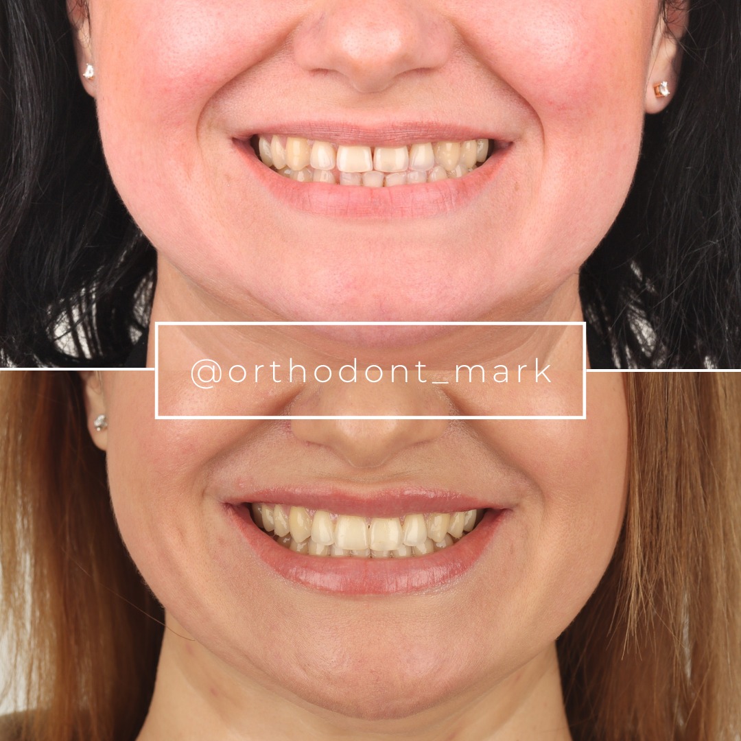
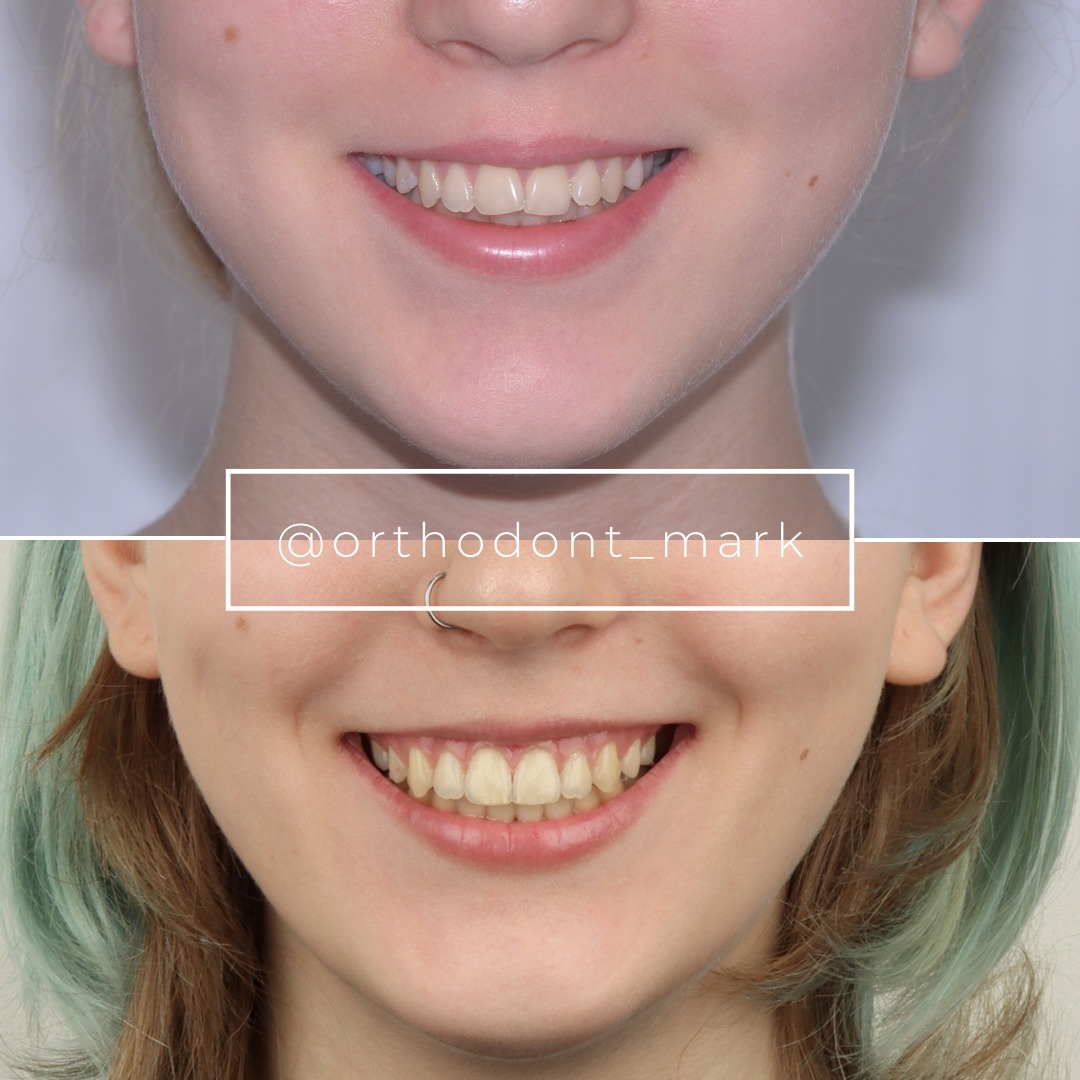
Clinical cases from Dr. Mark Radzhabov's practice
Learn the full MARPE protocol from Dr. Mark Radzhabov
Fundamental course covering CBCT patient selection, miniscrew planning, activation protocols, and 60+ clinical cases. Choose the access level that fits your practice.
Essentials of rapid palatal expansion for practicing orthodontists.
- Core RPE concepts and biomechanics
- 6 structured video lessons
- Clinical decision checklists
- Lifetime access to recordings
Deep-dive into MARPE protocol, diagnostics, and clinical execution.
- 6 modules covering MARPE from A to Z
- CBCT case selection walkthroughs
- Miniscrew placement and activation
- 60+ annotated clinical cases
- Direct Q&A with Dr. Mark Radzhabov
5-element medical consultation framework for dentists and orthodontists.
- Trust-building consultation protocol
- 5 lesson modules
- Templates for treatment plan delivery
- Works with any clinical specialty
Clinical FAQ
When is the optimal timing to begin facemask protraction after MARPE in late adolescents with Class III constriction?
Protraction should begin only after radiographic confirmation of midpalatal suture separation, resolution of peri-miniscrew gingival inflammation, and normalization of expansion-phase pain. Chun et al. (2022) reported separation in 95% of MARPE patients, but imaging confirmation remains the protocol gate, not elapsed activation time.
Does sex influence MARPE suture separation success in late adolescents undergoing combined expansion-protraction?
Yes. Jeon et al. (2022) found that older age significantly increased suture separation failure risk in males but not females. Clinicians should stratify male and female late adolescents separately when selecting candidates for the combined MARPE and facemask protraction protocol.
How common is asymmetric expansion in MARPE patients and how does it affect protraction sequencing?
Yoon et al. (2022) reported asymmetric expansion exceeding 1 mm in 47.8% of MARPE patients. Bilateral asymmetry creates an unequal force platform for subsequent protraction, making CBCT-based symmetry assessment a necessary checkpoint before facemask activation.
What is the prevalence of gingival inflammation around MARPE miniscrews and how should it be managed before protraction?
Yoon et al. (2022) identified gingival inflammation as the most frequent MARPE complication, affecting 83.9% of patients. Soft-tissue health should be established before protraction forces are introduced, as adding orthopedic load to inflamed peri-miniscrew tissue may worsen patient compliance.
Does MARPE produce greater maxillary skeletal width than conventional RPE as a foundation for facemask protraction?
Chun et al. (2022) demonstrated that MARPE produced greater maxillary width at the premolar and molar regions compared to RPE, suggesting bone-borne mechanics create a broader and potentially more stable skeletal base for subsequent protraction in Class III constriction cases.
How should clinicians counsel older male late adolescents about MARPE failure risk before beginning a combined protraction protocol?
Jeon et al. (2022) found age-related suture separation failure specifically in males. Consent documentation for older male adolescents should acknowledge the elevated expansion failure risk and establish a clear decision pathway — including surgical alternatives — if separation is not achieved.
What percentage of MARPE patients report pain during expansion and how does that affect facemask wear compliance?
Yoon et al. (2022) found that 45% of MARPE patients reported pain during or after expansion. Since facemask protraction requires consistent daily wear, introducing traction before expansion-phase discomfort has resolved is likely to reduce effective wear hours and compromise sagittal outcomes.
Is CBCT mandatory before initiating facemask protraction following MARPE in a late adolescent Class III case?
Evidence supports CBCT evaluation given that nearly half of MARPE patients develop asymmetric expansion exceeding 1 mm (Yoon et al., 2022). Bilateral comparison of palatal vault widening at the molar level confirms whether the skeletal foundation for protraction is symmetric before traction is applied.
How does MARPE suture separation rate compare with RPE in the context of combined expansion-protraction for Class III patients?
Chun et al. (2022) reported suture separation in 95% of MARPE patients versus 90% in RPE patients. The higher rate with bone-borne mechanics supports MARPE as the preferred expansion modality when reliable suture separation is a prerequisite for subsequent facemask protraction.
What imaging criteria should confirm suture separation before facemask protraction begins in class III constriction correction?
A visible midpalatal radiolucency on periapical or CBCT imaging, consistent with sutural diastema formation, is the standard radiographic indicator. Clinicians should verify this bilaterally and assess expansion symmetry before transitioning to the protraction phase of the combined protocol.
The evidence reviewed here reinforces a clear hierarchy: confirm suture separation before loading protraction forces, monitor soft-tissue complications weekly, and account for sex-based differences in suture response when selecting candidates. Late adolescents are not a homogeneous group, and protocol rigidity costs outcomes. Dr. Mark Radzhabov encourages clinicians to submit a challenging Class III case for review at ortodontmark.com, or to enrol in structured MARPE training to build the imaging and activation skills this combined protocol demands. Key sources: Chun et al. (2022), BMC Oral Health, doi:https://doi.org/10.1186/s12903–022-02138-w. Jeon et al. (2022), Clinical Oral Investigations, doi:https://doi.org/10.1007/s00784–021-04281–0. Yoon et al. (2022), retrospective analysis.

