
RPE Arch Perimeter Space Gain Basics
A Clinician's Guide to Expansion Mechanics in Growing Patients
Evidence-based breakdown of suture separation, device force output, and age-dependent expansion geometry for orthodontists planning transverse correction.
TL;DR Understanding rpe arch perimeter space gain basics helps clinicians select devices, time treatment, and anticipate skeletal versus dental outcomes in growing patients. Expansion force ranges, suture biology, and age-dependent opening geometry all interact. Bone-borne anchorage reduces dental tipping while preserving sutural separation. Matching device to patient age and suture maturity remains the central clinical decision.
A ten-year-old presenting with a posterior crossbite and moderate crowding forces a rapid decision: which expander design, what force level, and how will the dental arch perimeter actually change? Understanding rpe arch perimeter space gain basics—the mechanics linking transverse expansion to usable arch length—is non-negotiable for evidence-based case planning. At ortodontmark.com, Dr. Mark Radzhabov distills the published biomechanical and histological evidence so clinicians can move from radiographic diagnosis to protocol selection with confidence, without relying on convention alone.
How Does Sutural Opening Actually Translate to
Arch Perimeter
Gain in Growing Patients?
Arch perimeter space gain from rapid palatal expansion is the increase in available dental arch length that results when transverse maxillary widening separates the midpalatal suture and reshapes the alveolar arc. The clinical value of understanding this mechanism is that it prevents over-reliance on dental tipping as a substitute for true skeletal widening. When the suture parts, each half of the maxilla displaces laterally, adding circumferential length to the arch and relieving anterior crowding without extracting teeth. Suture separation is the key biological event, and anchorage design directly influences how reliably it occurs. Chun et al. (2022) compared conventional tooth-borne RPE against bone-borne MARPE in a prospective randomized clinical trial: midpalatal suture separation occurred in 90% of RPE patients and 95% of MARPE patients in their respective groups of 19 and 20 subjects. The difference in separation rate reflects anchorage geometry rather than activation force alone—bone-borne vectors distribute load more directly through palatal cortical bone toward the suture. The histological sequence following separation matters for consolidation timing. Caprioglio et al. (2017) collected midpalatal suture biopsies at 7 and 30 days post-expansion and documented active cellular remodeling at both intervals. By 30 days, newly formed bone trabeculae oriented perpendicular to the suture axis were considerably more prominent than at 7 days—confirming that the consolidation window requires weeks, not days, of appliance retention before the newly gained arch perimeter stabilizes. Removing the expander prematurely risks losing the transverse dimension before ossification is complete.
Which Expansion Screw Force Range Drives
Reliable
Sutural Separation Without Overloading Anchor Teeth?
Choosing between expansion screw designs is not an aesthetic preference. It is a biomechanical decision. Camporesi et al. (2013) used an Instron machine with 10 repetitions per screw type to measure forces at 1, 5, 10, 15, and 20 quarter-turns across three device types. The Hyrax and A2620 expanders generated forces exceeding 20 kg, while Palatal Split screws generated approximately 16 kg—with statistically significant differences between device types. All three fell within a force range adequate for rapid maxillary expansion, but the absolute magnitudes differ in ways that affect anchor tooth stress. Activation protocol must be calibrated to the device's force curve. A screw that reaches peak force early in its turn sequence loads the suture differently than one whose force climbs progressively. Clinicians prescribing one-turn-per-day schedules for a high-force Hyrax are delivering substantially greater instantaneous loads than they would with a Palatal Split design—a distinction that becomes clinically meaningful when periodontal support is limited or when the patient is near the upper age boundary for predictable suture opening. For structured RPE training for growing patients that addresses device selection alongside activation protocols, ortodontmark.com provides a dedicated curriculum. The practical implication is straightforward: confirm the force output characteristics of the specific screw you stock before writing an activation schedule, rather than importing a protocol from a device you no longer use.
Does Expansion Geometry Change the Net
Arch Length
Gain Across Age Groups?
The spatial pattern of maxillary widening is not fixed across development. Kinzinger et al. (2022) performed dental cast analysis on 60 growing patients stratified into three age groups of 20 each: under 10 years, 10–12 years, and 12 years and older. The analysis revealed that expansion in children under 10 years proceeds in a relatively parallel pattern across the arch, whereas in adolescents aged 12 and older, the opening becomes V-shaped, with anterior predominance. This shift has direct consequences for arch perimeter calculations: an equal screw activation does not produce equal anterior space gain across age groups. The V-shaped pattern in older adolescents means that posterior transverse gain at the molar region may exceed anterior gain. Clinicians planning to relieve anterior crowding through transverse expansion in patients aged 12 and older should anticipate that a disproportionate share of screw activation translates to posterior width rather than anterior arch perimeter—which may require adjunctive mechanics or modified activation schedules. This is not a failure of the appliance. It is an anatomical reality documented across a stratified sample. Beyond dental cast geometry, skeletal effects also differ by anchorage type. Chun et al. (2022) reported that MARPE produced significantly greater increases in nasal width at the molar region and at the greater palatine foramen compared to conventional RPE during both expansion and consolidation phases. These nasal and skeletal changes compound the arch perimeter gain by widening the basal bone support beneath the expanded dental arch.
How Should Clinicians Select Between Bone-Borne and Tooth-Borne
Expansion
When Planning Arch Perimeter Outcomes?
Device selection in growing patients should flow from two parallel assessments: the expected expansion geometry given the patient's age, and the tolerance of anchor teeth for lateral tipping forces. For patients under 10, tooth-borne RPE delivers adequate sutural force within the documented 16–20 kg range per Camporesi et al. (2013), and the parallel opening geometry means anterior arch perimeter gain is predictable. For patients approaching or past age 12, the V-shaped opening pattern documented by Kinzinger et al. (2022) reduces anterior efficiency, and any tendency toward anchor tooth buccal displacement—measurable and significant per Chun et al. (2022)—amplifies dental side effects without adding skeletal width. Bone-borne designs shift the force vector away from anchor teeth entirely, delivering load through miniscrews into palatal cortical bone. This is particularly relevant when periodontal bone height is reduced, crown-to-root ratios are unfavorable, or when the clinician needs skeletal nasal base widening in addition to dental arch perimeter gain. The nasal and greater palatine foramen changes documented by Chun et al. (2022) for MARPE versus RPE confirm that the skeletal envelope of expansion is genuinely broader with bone-borne anchorage. Consolidation management deserves equal protocol attention. Given that Caprioglio et al. (2017) documented active trabecular remodeling persisting through 30 days, early appliance removal before this window closes undermines the very arch perimeter gain the expansion achieved. Retention duration should be driven by the histological timeline, not by patient comfort or scheduling convenience. The MARPE skeletal expansion protocol page at ortodontmark.com details consolidation timing alongside activation schedules grounded in this evidence.
Free Clinical Guide
Which Expansion Appliance at Which Age?
Get Dr. Mark Radzhabov's guide to skeletal-expansion appliances — indications, pros & cons, and how to choose by patient age. Free to your inbox.
Prefer video? Watch the free demo lesson on MARPE appliance types, features and trade-offs.
Documented outcomes
Clinical cases from Dr. Mark Radzhabov's practice
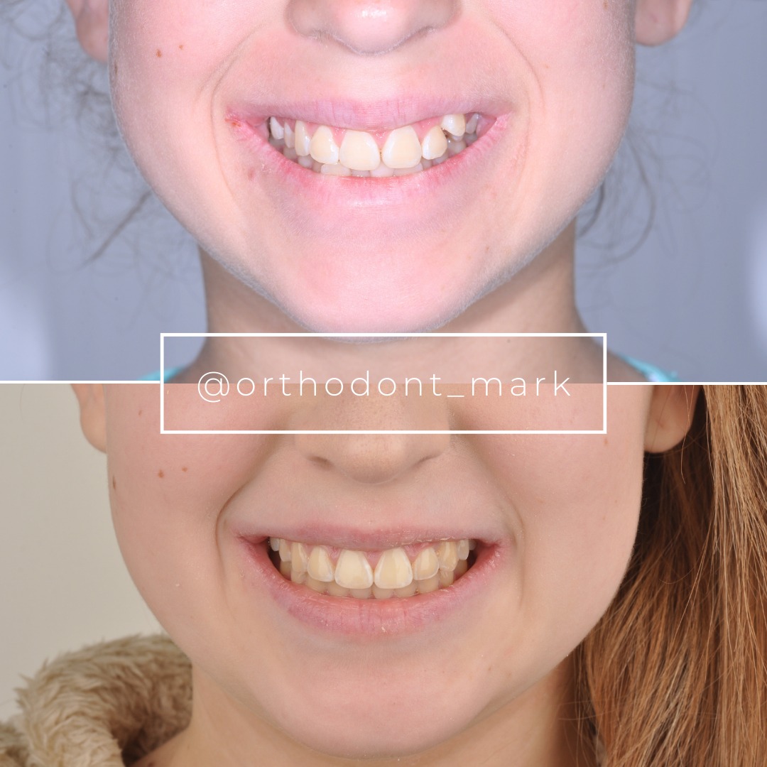
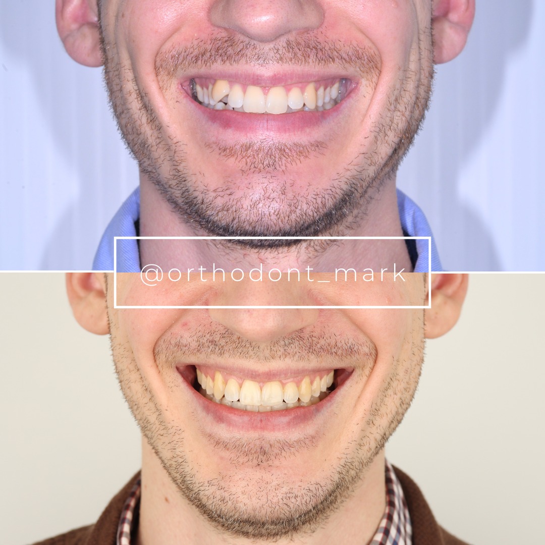
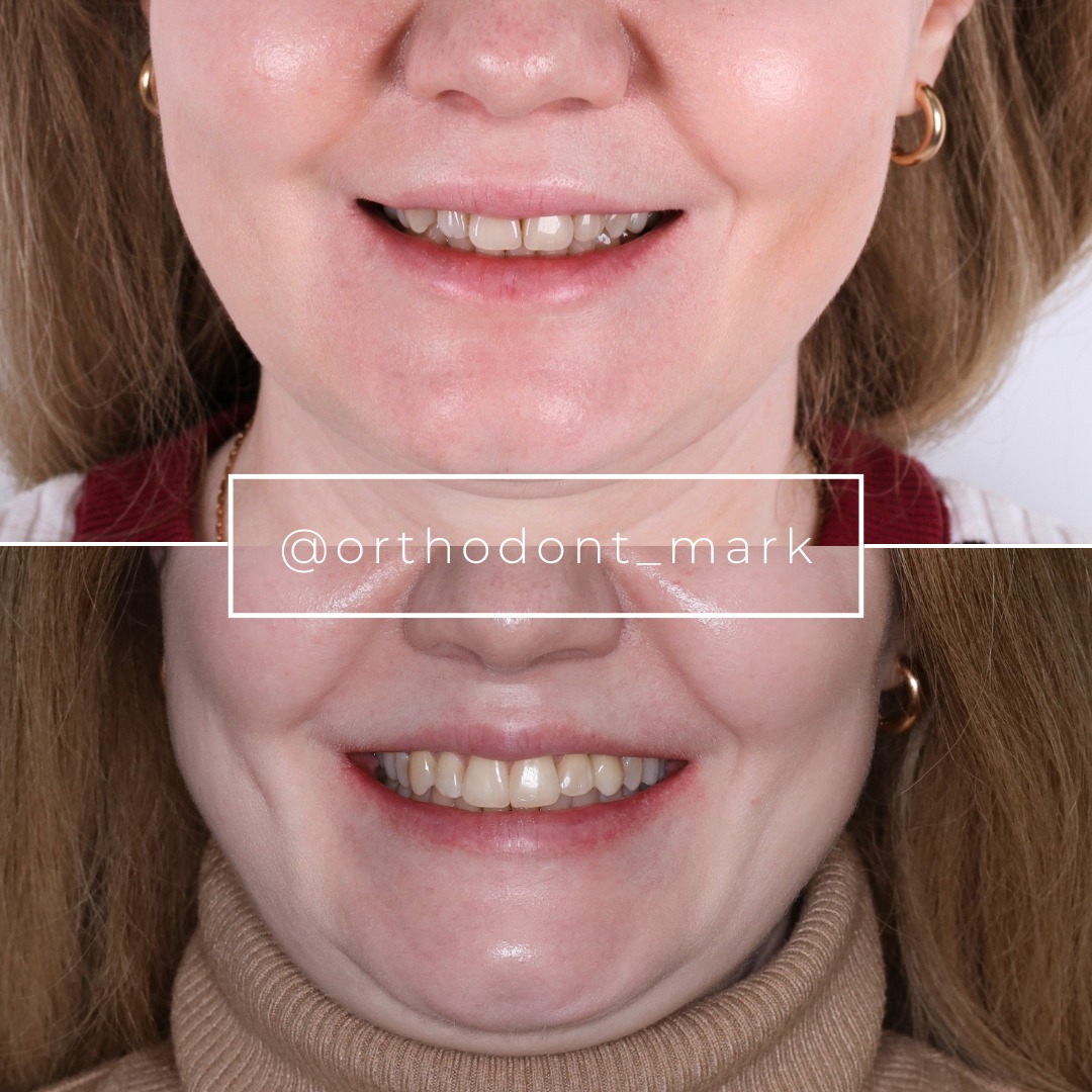
Learn the full MARPE protocol from Dr. Mark Radzhabov
Fundamental course covering CBCT patient selection, miniscrew planning, activation protocols, and 60+ clinical cases. Choose the access level that fits your practice.
Essentials of rapid palatal expansion for practicing orthodontists.
- Core RPE concepts and biomechanics
- 6 structured video lessons
- Clinical decision checklists
- Lifetime access to recordings
Deep-dive into MARPE protocol, diagnostics, and clinical execution.
- 6 modules covering MARPE from A to Z
- CBCT case selection walkthroughs
- Miniscrew placement and activation
- 60+ annotated clinical cases
- Direct Q&A with Dr. Mark Radzhabov
5-element medical consultation framework for dentists and orthodontists.
- Trust-building consultation protocol
- 5 lesson modules
- Templates for treatment plan delivery
- Works with any clinical specialty
Clinical FAQ
How does rapid palatal expansion increase dental arch perimeter in growing patients?
Midpalatal suture separation displaces each maxillary half laterally, adding circumferential length to the alveolar arc. This transverse gain converts to anterior arch perimeter, reducing crowding without extraction. The conversion efficiency depends on patient age and the expansion geometry produced.
What age-related change in expansion geometry affects anterior arch length gain from RPE?
Kinzinger et al. (2022) documented a shift from parallel expansion in children under 10 years to a V-shaped, anteriorly predominant pattern in adolescents aged 12 and older. In older patients, more screw activation reaches the posterior arch, reducing anterior space gain per unit of activation.
What force range do common expansion screws produce during rapid palatal expansion?
Camporesi et al. (2013) measured Hyrax and A2620 expanders exceeding 20 kg and Palatal Split screws generating approximately 16 kg across 20 quarter-turns in standardized Instron testing. All three device types fell within the force range considered adequate for rapid maxillary expansion.
How long should a palatal expander be retained to consolidate arch perimeter space gain?
Caprioglio et al. (2017) biopsied the midpalatal suture at 7 and 30 days post-expansion. Bone trabeculae perpendicular to the suture axis became significantly more prominent by 30 days. Retention should span at least this biological window before appliance removal is considered.
How does anchor tooth buccal displacement differ between RPE and MARPE in growing patients?
Chun et al. (2022) found MARPE produced significantly less buccal displacement of anchor teeth compared to conventional RPE during both expansion and consolidation. Bone-borne anchorage directs force through palatal cortical bone rather than through the crowns of anchor premolars or molars.
Does bone-borne MARPE produce different skeletal nasal effects than tooth-borne RPE?
Yes. Chun et al. (2022) reported MARPE produced significantly greater increases in nasal width at the molar region and at the greater palatine foramen compared to RPE. These basal skeletal changes extend beyond the dental arch and support the expanded alveolar perimeter more broadly.
How does suture separation rate compare between tooth-borne and bone-borne expanders?
Chun et al. (2022) documented midpalatal suture separation in 90% of RPE patients and 95% of MARPE patients. Both figures are high, but anchorage design influences the reliability and the dental side-effect profile alongside the separation outcome.
How should clinicians counsel patients on the palatal expansion consolidation timeline?
Inform patients that the expander must remain in place during active bone deposition across the opened suture. Histological data from Caprioglio et al. (2017) confirm meaningful trabecular formation continues through at least 30 days, so early removal is a biological risk, not merely a protocol deviation.
At what age does expansion geometry shift from parallel to V-shaped, and why does it matter for clinical planning?
Kinzinger et al. (2022) identified the shift occurring at age 12 and older. The V-shaped pattern concentrates width gain posteriorly, so clinicians planning to relieve anterior crowding must adjust activation goals or add anterior arch mechanics to achieve the targeted arch perimeter increase.
How does screw type selection affect the clinical protocol for rapid palatal expansion?
Different screws reach different force plateaus. Camporesi et al. (2013) found significant differences between device types in the 16–20 kg range. Importing an activation schedule calibrated to one screw design and applying it to a higher-force device risks overloading anchor teeth and sutural structures.
Transverse expansion in growing patients is mechanically straightforward only when device selection, activation force, and patient age are matched deliberately. The evidence reviewed here—covering suture biology, force output variability, and age-dependent opening geometry—gives clinicians a concrete framework rather than a rule of thumb. Reviewing your case mix against these findings is a practical next step. Dr. Mark Radzhabov offers structured guidance through ortodontmark.com, where dedicated RPE and MARPE courses translate this evidence directly into clinical workflow and consent protocols.